Researchers rolled up their sleeves and produced important analyses of the Comparison of Age-Related Macular Degeneration Treatment Trial (CATT) and the MARINA and ANCHOR studies. VEGF Trap-Eye, sleep apnea, and other topics also stimulated rich discussion of future interventions in the posterior segment.
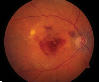
Research at ARVO followed up on the CATT study, which confirmed that Avastin is just as effective as Lucentis for the treatment of neovascular AMD, as seen in this patient. In this new research, however, clinicians are starting to see post-treatment differences in outcome.
Milestone Trial Continues Its Influence
Following up on the landmark findings of CATT, researchers confirmed that the reduction of central neovascular (CNV) lesion sizes after injection with ranibizumab was similar to the reduction experienced after injection with bevacizumab.2893/A338 However, they were also starting to see post-treatment differences in outcome that were determined by retinal anatomy.
In a review of 1,185 patients after 52 weeks of treatment, the researchers found anti-vascular endothelial growth factor (anti-VEGF) therapy reduced lesion activity and improved visual acuity in all treatment groups. However, at all time points, those with residual OCT-determined intraretinal fluid had worse visual acuity than those without fluid. In addition, abnormally thin or thick retinas revealed worse visual acuity. Monthly ranibizumab dosing resulted in more eyes with no fluid and lower mean retinal thickness, although the long-term significance of this finding was still unknown. These results raised important treatment implications in eyes undergoing anti-VEGF therapy for neovascular age-related macular degeneration (AMD).
In another CATT follow-up report, anti-VEGF agents were found to possibly have a systemic impact on the proliferation of AMD in the fellow, unaffected eye.3680 This is important data for optometrists, helping to inform their efforts in continuously monitoring for the appearance or progression of disease in contralateral eyes.
Among 1,185 CATT patients, 61% showed no signs of CNV in the fellow eye at enrollment. At one year, CNV had developed in 8% of 365 eyes of patients who had been treated with ranibizumab and seven percent of 362 patients who had been treated with bevacizumab. After adjusting for known risk factors for CNV and drug dosing regimen, the estimated hazard ratio associated with treatment for bevacizumab was 0.92 (95% confidence interval [0.54, 1.56]). As a result, the authors concluded that CNV incidence rates in the fellow eyes of patients treated with anti-VEGF agents could indicate the systemic effects of the drugs. Through one year, ranibizumab and bevacizumab had similar effects on the incidence of CNV in the fellow eye.
Genetic Implications
Another follow-up on CATT looked at response to anti-VEGF agents among patients who had primary genetic factors for AMD.3682 The response in these patients was virtually identical to the response in patients who were not genetically predisposed to AMD. To reach this conclusion, researchers evaluated 75% of 1,116 patients participating in CATT at 43 clinical centers. Each patient was genotyped for single nucleotide polymorphisms (SNPs) rs1061170 (CFH), rs10490924 (ARMS2), rs11200638 (HTRA1), and rs2230199 (C3), using TaqMan SNP genotyping assays. These results provided long-term hope for the visual outcome of patients with CFH2 or ARMS2 genetic variants.
Despite this promising conclusion, another study offered a troubling and completely opposite finding.3683 Researchers in this case determined that genetically predisposed patients experienced onset of AMD at an earlier age and often responded poorly to treatment with ranibizumab, compared to patients without the genetic variants.3683
Researchers evaluated 420 eyes of 397 unrelated Caucasian neovascular AMD patients who had been treated only with intravitreal 0.5mg ranibizumab injections. Each participant underwent best-corrected visual acuity testing before and after treatment with three ranibizumab injections. Genotyping of SNPs in the CFH, ARMS2, VEGF, kinase insert domain receptor (KDR), low-density lipoprotein receptor-related protein 5 (LPR5) and FZD4 was performed.
In addition, a very important study showcased early genetic treatment for patients with Leber congenital amaurosis.4642 Most patients experienced long-term visual gains with the use of an experimental oral therapy, QLT091001. The therapy was used for patients whose condition was associated with lecithin/retinol acyltransferase (LRAT) or retinal pigment epithelial 65 protein (RPE65) mutations.
Trap-Eye Data Released
Pivotal data from the latest anti-VEGF Trap-Eye study were released. These results suggest that every-other-month dosing of intravitreal aflibercept (Eylea, Regeneron Pharmaceuticals) is just as effective as monthly injections of ranibizumab (Lucentis, Genentech/Roche).2042/D1060
A total of 2,457 patients with AMD from VIEW 1 and VIEW 2 were randomized to four treatment groups: ranibizumab 0.5mg every four weeks and aflibercept 2mg every four weeks, aflibercept 0.5mg every four weeks, and aflibercept 2mg every eight weeks (after three initial monthly doses).
What is VEGF Trap-Eye?
On June 17, the FDA’s Dermatologic and
Ophthalmic Drugs Advisory Committee voted unanimously to recommend
approval of Eylea (aflibercept ophthalmic solution, Regeneron
Pharmaceuticals)—an injectible drug for the treatment of the neovascular
form of age-related macular degeneration, or wet AMD.
Eylea, also known
as VEGF Trap-Eye, is a fully human fusion protein consisting of
portions of VEGF receptors 1 and 2 that bind all forms of VEGF-A, along
with the related Placental Growth Factor (PlGF). Eylea is a specific and
highly potent blocker of these growth factors. The drug is specially
purified and contains iso-osmotic buffer concentrations, allowing for
injection into the eye.
In outcomes of ≥ 15 letters at the 52nd week, cumulative incidence curves for the four dosing regimens did not differ. Improvement in vision was observed early in all treatment groups.
Another VEGF Trap-Eye study examined the safety and efficacy of the drug for treating cystoid macular edema secondary to central retinal vein occlusion (CRVO).6929 The one-year GALILEO study—a double-masked, multi-center, controlled phase III study—looked at 177 patients who were randomized to 2mg of intravitreal aflibercept or sham injections every four weeks. Beginning at week 24 through week 52, patients in the aflibercept group were treated on an as-needed (PRN) basis with sham injections during non-treatment visits.
After 52 weeks, 60.2% of patients in the aflibercept group had gained at least 15 ETDRS letters from baseline, compared to 32.4% of patients in the sham group. These results corroborated previous results from the sister COPERNICUS study, suggesting that intravitreal aflibercept injection could be an effective treatment for macular edema secondary to CRVO.
Long-term MARINA and ANCHOR Results
Seven-year data from MARINA and ANCHOR were released, suggesting that most patients on long-term anti-VEGF therapy continued to demonstrate significant disease progress or blindness.3679
Fourteen U.S. clinical trial sites recruited 65 patients originally treated in the ANCHOR and MARINA trials (enrolled between March 2003 and September 2004) and further treated with ranibizumab in the HORIZON extension study. At the time of this most recent analysis, the cohort was being reviewed seven to eight years after initiation of intravitreal ranibizumab treatment. For the primary endpoint, 37% of original study eyes had ETDRS visual acuity of 20/70 or better. The mean visual acuity in study eyes was 20/125. Within the cohort, subgroups showed favorable outcomes, with good vision (≥ 20/40) in 23% of eyes and durable CNV quiescence in 35%. By contrast, another group showed poor outcomes; 37% of eyes had vision of 20/200 or worse. Ongoing exudative disease activity, defined as evidence of CNV leakage or hemorrhage at study visit or within the previous 6 months, was found in 54% of study eyes, and 23% required ongoing treatment (ranibizumab or other AMD treatments).
A report on the COMPLETE study, representing extremely important early work on a complement-inhibitor drug, was released. Complement-inhibiting agents represent the newest and most significant therapeutic treatments for early AMD.2045/D1063
In this prospective, double-masked study, patients with drusen in the absence of geographic atrophy were randomized 2:1 to intravenous (IV) eculizumab or placebo in a double- masked fashion. Fifty percent of patients in the eculizumab group received a low-dose regimen of 600mg weekly for four weeks, followed by a maintenance period of 900mg every two weeks until week 26. The other 50% received a high dose of 900mg weekly for four weeks, followed by 1,200mg every two weeks until week 26.
The COMPLETE study represents the first coordinated use of systemic complement inhibition for the treatment of dry AMD and systemic complement inhibition. Eculizumab was well tolerated through six months.
Sleep Apnea and Bevacizumab
Researchers found that untreated obstructive sleep apnea hinders functional and anatomical response to bevacizumab in AMD.2925/A370 The treatment of sleep apnea is a popular topic across multiple disciplines of medicine, particularly in eye care (glaucoma, ocular surface disease and, now, AMD). These results suggest that optometrists should become more knowledgeable about sleep apnea, the overall impact of the condition and how to treat and manage it with other healthcare professionals.
Twelve patients with untreated obstructive sleep apnea were treated with intravitreal bevacizumab (1.25mg/0.05ml) injections every six weeks for clinical and angiographic evidence of exudative AMD. Clinical examinations and OCT every six weeks were used to assess the anatomical and functional outcome for up to 90 weeks. The treatment of obstructive sleep apnea with continuous positive airway pressure yielded a subsequent impressive anatomical response, but functional improvement did not follow. Identifying and treating underlying obstructive sleep apnea earlier in the management of exudative AMD may provide better functional outcomes.
New Approaches to Retinal-vitreal Disease
A new treatment for retinal-vitreal disease was shown to be well tolerated.1337 The data suggested promising potential, despite the study’s limited size. ALG-1001 is a synthetic anti-integrin oligopeptide. The first human clinical safety and efficacy data on this new class of anti-angiogenic compounds in the eye was presented.
Studies to date have shown that ALG-1001 inhibits integrin receptors in vitro and, in vivo, arrests aberrant blood vessel growth meditated by αvß3, αvß5, α2ß1 and α5ß1. These are integrin sites that are expressed in neovascular ocular tissue in patients with wet AMD and diabetic retinopathy.
Fifteen patients with end-stage diabetic macular edema completed this open label study. Baseline best- corrected visual acuity (BCVA) was ≥ 20/100. Patients had not undergone anti-VEGF treatment or focal laser treatment within 90 days. Many were refractory to bevacizumab and previous photocoagulation. Despite the small study size, the results demonstrated clinically significant efficacy, including improvements in BCVA and OCT-measured central macular thickness. The clinical improvements endured to the end of the study, at least 90 days past the last intravitreal treatment in nearly all study subjects who demonstrated improvements.
Ocriplasmin Moves Forward

Ocriplasmin could provide a minimally invasive pharmacologic approach to treat patients like this one who have VMT syndrome.
Phase III of the MIVI-TRUST study provided further evidence that ocriplasmin could effectively remove vitreomacular adhesion (VMA) as well asfoster the repair of full-thickness macular holes without surgical intervention.2754 Also, the treatment appears both safe and well tolerated.
The MIVI-TRUST program, which consisted of two large, phase III clinical trials, investigated a single intravitreal injection, 125µg (100µl) of ocriplasmin, compared to a single 100µl placebo injection for the pharmacological treatment of symptomatic VMA.
A single intravitreal dose of 125µg of ocriplasmin achieved resolution of VMA in approximately 30% of patients. Resolution of this anatomic pathology resulted in clinically significant visual acuity benefits in patients with vitreomacular traction (VMT) syndrome. Treatment was well tolerated by patients. Ocriplasmin could provide a minimally invasive pharmacologic approach to treat patients with VMT syndrome.
No Sx Required for VMT?
A report on 36 eyes from Bascom Palmer suggested that patients with mild to moderate VMT syndrome did not require surgical intervention.5220/D1273 The clinical course of VMT for eyes that did not undergo surgery was relatively stable over 16 months of mean follow-up. Visual acuities at the initial and final follow-up visits were similar. The study identified a low rate of progression from mild to severe grades of VMT that required vitreoretinal surgery.
Comparing Technologies
A study that compared SD-OCT, camera-based fundus autofluorescence (AF), confocal scanning laser ophthalmoscope AF and fluorescein angiographic (FA) imaging raised potentially lucrative implications for imaging device companies. A total of 30 patients were enrolled in a geographic atrophy (GA) cohort. While GA can be measured using different imaging modalities, each modality measures a different property of GA. On average, the comparison found that areas of GA measured with AF and FA were smaller than the areas measured by SD-OCT images. Whether these different imaging modalities yield similar enlargement rates remained to be determined.
These results undoubtedly will be scrutinized by industry in search of breakthrough diagnostic and monitoring technology. Nonetheless, current data continues to suggest that the use of multiple imaging devices will likely be more accurate and clinically useful than relying on one single modality.
Positive long-term results were reported for Second Sight’s Argus II, a retinal prosthesis.6953 All 30 patients, who had bare light perception or worse because of retinitis pigmentosa, received Second Sight Argus II implants.
New Ways of Monitoring
Advances in treatments for AMD and diabetic retinopathy suggest that frequent disease monitoring is crucial for timely intervention. Recently, a new handheld shape discrimination hyperacuity (hSDH) test iPhone app was designed for visual function self-monitoring in patients with maculopathy.2914/A359
This appears to be a promising app for at-home monitoring. More apps are becoming available to provide useful clinical information to the user. This app could help patients determine if their condition is progressing, and persuade them to schedule an appointment for a clinical assessment.
