 History
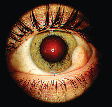
A 23-year-old white female presented for a routine eye examination. The patient said that her previous eye doctors were unable to explain the nature of a fleshy lump on the white of her left eye. She said that the lump had been there all her life, was not changing in size and (to the best of her knowledge) was a birthmark. She had no known allergies and denied the use of any medications.
Diagnostic Data
This 23-year-old female presented with a fleshy lump O.S. that she said was there her entire life. What caused the lump and will it negatively impact her vision in the future?
Her best-corrected entering visual acuity was 20/20 O.U. The pertinent anterior segment finding O.S. is illustrated in the photograph. The lesion was soft, symmetric, evenly colored, mobile and approximately 6mm in diameter.
Otherwise, her visual fields, refraction, biomicroscopy, tonometry and dilated fundus evaluation were unremarkable and noncontributory.
Your Diagnosis
How would you approach this case? Does this patient require any additional tests? What is your diagnosis? How would you manage this patient? Whats the likely prognosis?
Discussion
Additional testing should include palpation to ensure that the lesion is both soft and moveable. Also, be sure to check marginal reflex distance and exophthalmometry to rule out proptosis and intraorbital involvement. Neuroimaging is another available option for detection and diagnosis.
The diagnosis in this case is congenital conjunctival limbal dermoid. Conjunctival limbal dermoids are lined by nonkeratinized conjunctival epithelium with goblet cells and appear as firm, subcutaneous masses. They sometimes contain dermal appendages, such as hair shafts, sebaceous glands and sweat glands. Conjunctival limbal dermoids account for approximately 5% of dermoid cysts.1 While they are often congenital, conjunctival limbal dermoids are often not apparent until later in childhood or early adulthood.1
Cysts of the surface epithelium are divided into two categories: simple epithelial cysts (epidermal, conjunctival, respiratory and apocrine gland) and dermoid cysts (epidermal and conjunctival). Epidermal dermoid cysts are by far the most common orbital cystic lesion found in children; they account for more than 40% of all childhood orbital lesions and 89% of all childhood orbital cystic lesions that are biopsied or surgically removed.1
Goldenhars syndrome (oculo-auriculo-vertebral dysplasia) encompasses a wide spectrum of congenital anomalies involving structures that arise from the first and second branchial arches, including anophthalmos, lipodermoid, preauricular appendages and mandibular hypoplasia.1 Rule out a diagnosis of Goldenhars syndrome in patients who present with a limbal dermoid expression.
The majority of child-age patients who have had dermoid cysts surgically removed report excellent visual results.2 In most cases, younger children with Goldenhars syndrome or an episodic demoid cyst undergo corrective surgical procedures for visual reasons. However, these individuals often require larger grafts and have a tendency for lower visual acuity secondary to amblyopia of disuse. In these cases, the risks associated with a complicated surgical correction may outweigh the risks of waiting until the end of the childs sensitive amblyogenic period.3
In this case, our patient was presented with several corrective surgical options, but she declined all procedures.
1. Shields JA, Shields CL. Orbital cysts of childhoodclassification, clinical features and management. Surv Ophthalmol 2004 May-Jun;49(3):281-99.
2. Watts P, Michaeli-Cohen A, Abdolell M, Rootman D. Outcome of lamellar keratoplasty for limbal dermoids in children. J AAPOS 2002 Aug;6(4):209-15.
3. Berker N, Acaroglu G, Soykan E. Goldenhars syndrome (oculo-auriculo-vertebral dysplasia) with congenital facial nerve palsy. Yonsei Med J 2004 Feb 29;45(1):157-60.
