 |  |
Various eyelid lesions are a common occurrence among all patient populations, and their causes are numerous, with the vast majority being benign. Clinicians should always identify the etiology of the lesion—whether it’s inflammatory, infectious, structural or neoplastic—as it will direct the proper management.
When a benign neoplasm, such as a papilloma, is present on the eyelid margin, a shave excision is often the most appropriate course of action. Although most do not pose any visual compromise, the lesions can cause irritation such as foreign body sensation. Patients presenting with complaints of eyelid lesions are often looking for a cosmetic as well as therapeutic intervention. Clinicians should not dismiss cosmetic concerns, as patients may feel self-conscious about their appearance, and removal can be pivotal to their quality of life.
Removing these lesions is a beneficial procedure for these patients and can be done in the office. Check with your state board for specific regulations regarding optometrists performing this procedure.
Rule Out Malignancy
While most lesions are benign and can simply be removed, each should be evaluated for the possibility of malignancy prior to removal. If a lesion is suspicious or of unknown etiology, refer to an ophthalmic surgeon for excision and biopsy to rule out malignancy. Signs of eyelid malignancy include madarosis and alterations to the normal eyelid integrity.1 Malignant lesions also tend to grow faster than those that are benign. They can be painful, bleed more easily and form ulcers or crusts in the center of the lesion.
Margin Integrity
When lesions are present on the eyelid margin, extra surgical care is warranted, as the structural integrity of the eyelid margin is essential to appearance and functionality. A shave excision is useful when biopsy is warranted, but a level of precision is necessary to maintain margin integrity. It provides the surgeon with histological control over the tissue being excised.
All excised tissue specimens should be sent to a pathology lab for histopathologic analysis. Results are usually returned in one to two weeks, after which you should follow up with the patient to discuss.
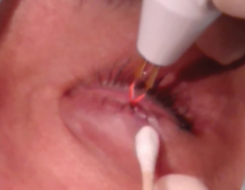 |
| After the excision, cauterize the wound to control any bleeding. |
Procedure Basics
Prior to excision, the lower eyelid should be anesthetized with lidocaine 2% with 1:100,000 epinephrine. This also induces vasoconstriction of the surrounding vessels, limiting blood loss following the procedure. The area is prepped with betadine, draped and tested for anesthetic effect.
After confirmation of anesthesia, the lesion is grasped with forceps, exposing its base. An 11-degree surgical blade is used to remove the lesion at its base on the lid margin. Care is taken to ensure no notches are made in the margin itself. The removed specimen is placed into container and medium supplied by the lab and sent for analysis.
Cautery helps to control bleeding, and ophthalmic antibiotic ointment is used to prevent infection.
Following the procedure, the patient is instructed to use antibiotic ointment twice a day for five days.
A simple procedure, shave excision can have a significant impact on patients who are encumbered by common eyelid lesions.
Dr. Skorin is a consultant in the Department of Surgery, Community Division of Ophthalmology in the Mayo Clinic Health System in Albert Lea, MN.
Dr. Goemann received her degree at Pacific University College of Optometry and practices at Eye Q Vision in Mankato, MN and Family Eye Care in Fairmont, MN.
| 1. Carter S. Eyelid disorders: Diagnosis and management. Am Fam Phys. 1998:2695-2702. |
