When a 36-year-old male patient came in for a routine MRI to track the progress of bilateral optic nerve sheath meningiomas, Jonathan C. Horton, MD, PhD, and Steven Miller, MD, PhD, found more than they expected.
The MRI scans revealed inflammation in the right orbit, prompting immediate follow up in the clinic. They found that the patient had developed epidemic keratoconjunctivitis (EKC) but had not complained of symptoms prior to the scheduled appointment. While inflammation is common on the conjunctival surface, this patient’s MRI revealed some interesting surprises below the surface.
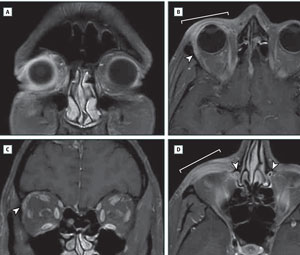 | |
| A. Fat saturation, demonstrating enhancement of periocular tissues. B. Thickening of the right upper eyelid (bracket) and anterior orbital enhancement. Lacrimal gland is enlarged (arrowhead). C. Right lacrimal gland enlargement (arrowhead). D. Inflammation of the lower eyelid (bracket) and right nasolacrimal duct swelling with loss of air signal in the lumen (arrowheads). |
“The infection induces an inflammatory process that extends surprisingly deep into the orbit,” the authors write. Published online May 28 in JAMA Ophthalmology, the primary findings based on the MRI imaging “were edema and inflammation of periocular tissue in the anterior orbit, enlargement of the lacrimal gland, and nasolacrimal duct compromise.”
“Our case presented as a typical infection and the patient recovered uneventfully,” the authors conclude, “indicating that deep tissue inflammation, dacryoadenitis and dacryocystitis are likely to be common manifestations of adenoviral conjunctivitis.”
“Clinicians should still rule out a bacterial cellulitis or preseptal cellulitis, but the most common diagnosis of a severe conjunctivitis presentation is epidemic keratoconjunctivitis, and that is now evident with these MRI images,” says Paul Karpecki, OD, head of the ocular surface disease clinic and director of clinical research at the Koffler Vision Group in Lexington, Ky.
This case study has implications for clinical practice, too. “It also shows the need for topical corticosteroids in severe cases given the level of inflammation that can be present,” Dr. Karpecki says. “Finally, it helps us empathize with patients with epidemic keratoconjunctivitis who are miserable, and now we may have good evidence to understand why.”
While Dr. Karpecki would love to see a larger study to confirm the findings, he notes MRI imaging can be cost prohibitive, and the data in this case can be sufficient to inform clinical decisions.
Horton J, Miller S. Research letter: Magnetic Resonance Imaging in Epidemic Adenoviral Keratoconjunctivitis. JAMA Ophthalmology. 2015 March 28. doi:10.1001/jamaophthalmol.2015.1457.