A 64-year-old white male presented with a three-week history of irritation and redness at the lid margins O.U. The patient was diagnosed with rosacea about three years earlier. At the time of examination, he was using topical metronidazole for associated skin lesions.
His best-corrected visual acuity was 20/20 O.U. Biomicroscopy revealed mild blepharitis and significant meibomian gland dysfunction (MGD) O.U. Mild, diffuse conjunctival hyperemia was also present O.U. A grade 1 punctate epithelial keratopathy was evident on the inferior aspect of each cornea, without infiltrate or neovascularization. All other ocular findings were normal O.U.
Based on the medical history and appearance, we diagnosed the patient with ocular rosacea. We initiated treatment with warm compresses applied to the eyelid margins t.i.d., non-preserved artificial tears q3h, and 100mg oral doxycycline b.i.d. for six weeks.
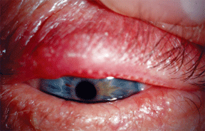
Up to 60% of individuals with rosacea will experience associated ocular manifestations. Note redness and swelling of the lid margins in this patient. Courtesy: Alan G. Kabat, O.D.
Additionally, we counseled the patient to avoid triggers, such as sunlight, spicy foods, hot beverages and stress. A follow-up examination four weeks later showed near complete resolution of associated signs and symptoms.
Currently, the patient has regular dermatology and optometry visits and remains on doxycycline monohydrate and topical skin therapy.
Rosacea affects about 14 million Americans.1 Approximately one in 20 adults in the
In this article we present a discussion on rosacea, with emphasis on both differential diagnosis and management of the ocular and underlying dermatologic pathology.
The Skinny on Skin
Skin is responsible for several functions. First, it protects the body from the surrounding environment; it actively prevents and combats infection. Also, skin helps regulate body temperature. Finally, skin is a sensory organ that detects temperature, touch, pain, vibration and other sensations. Skin is constantly working to renew itself.
The epidermis is the skins outermost layer. It consists of two main cell types: keratinocytes and melanocytes. These cells are produced in the basal layer of epidermis. The epidermis contains no blood vessels; however, it does have a protective outer layer, the stratum corneum.3
The dermis is beneath the epidermis and consists of a thicker layer of fibrous connective tissue. It supports and binds the epidermis to the subcutaneous tissue. The dermis produces collagen, elastin and reticulinall substances that lend structure and support. The dermis provides nutrition to both itself and the epidermis. The dermis contains nerves, blood vessels, lymph vessels and sebaceous glands.3
The hypodermis is a subcutaneous layer comprised of loose connective tissue. It also contains variable amounts of adipose. The hypodermis contains larger blood vessels and nerves than those found in the dermis. Fibrous bands in the hypodermis serve to anchor the skin to the deep fascia.3
Rosacea and its associated symptoms predominantly occur in four stages:5 Prerosacea. The hallmark of this early stage is flushing, or recurrent episodes of facial redness. Sunlight, alcohol, tobacco, spicy or hot foods and beverages, and stress commonly trigger flushing. The patient may develop erythema, a persistent mid-facial redness. The nose, chin, cheeks and central forehead may all be affected. Stage 1. This stage of the condition includes signs of prerosacea, plus telangiectasia, a permanent dilation of small blood vessels, and prominent sebaceous glands, resulting in an oily appearance to the skin. These early vascular signs occur in an axial facial distribution and may show a butterfly pattern of breakout across the nose and cheeks. Stage 2. This stage includes all potential signs of stage 1, plus edema of the involved tissue, papules (skin bumps) and pustules (pimples), enlarged pores and raised red patches (plaques). Stage 3. This stage includes all potential signs of stage 2, plus tissue hyperplasia (or phymas), and rhinophyma, or enlargement of the sebaceous glands of the nose.
Understanding Rosacea
Recognizing Rosacea
Rosacea is a chronic dermatologic condition that affects the convexities of the central aspect of the face, including ocular tissues. The condition is characterized by symptoms of facial flushing and a spectrum of clinical signs, such as erythema, telangiectasia, coarseness of the skin and an inflammatory papulopustular eruption that resembles acne.4
Rosacea is a clinical diagnosis that does not require laboratory testing or pathology specimens. Differential diagnoses for rosacea include acne vulgaris, contact dermatitis, seborrheic dermatitis, eczema, sarcoidosis, lupus, perioral dermatitis and drug-induced photosensitivity (i.e., the reaction from tetracycline).4
There are four distinct subtypes of rosacea.5 Many patients have characteristics of more than one subtype. They include:
Erythematotelangiectatic rosacea, which is characterized by blood vessel dilatation and telangiectatic changes.
Papulopustular rosacea, which is characterized by skin bumps, pimples and plaques.
Phymatous rosacea, which is characterized by phymas, or tissue hyperplasia.
Ocular rosacea, which is characterized by myriad ocular signs.
Rosacea is characterized by exacerbations and remissions. Treatment of rosacea empirically targets these signs and symptoms because investigators do not precisely understand the conditions pathophysiology. Long-term therapy is usually required to control the effects of rosacea.
Rosacea is more common in fair-skinned individuals, but is probably under-reported in ethnic populations with increased skin pigmentation. The peak incidence occurs during the fourth to seventh decades of life. Females are affected two to three times more often than males; however, the condition is often more severe in men.6 Rosacea commonly affects the cheeks in women and the nose in men.
Ocular Rosacea
Ocular signs and symptoms present before skin manifestations occur in up to 20% of patients who are ultimately diagnosed with rosacea. The primary ocular symptoms are foreign-body sensation, burning and stinging. The etiology of ocular rosacea is inflammation from Staphlococcus exotoxins.7 The ocular signs are secondary to the inflammatory skin condition.
Ocular surface inflammatory disease is the principal complication of the ocular rosacea subtype.8 Clinical signs include dry eye, telangiectasia of lid margins, conjunctivitis, blepharitis, recurrent chalazia and hordeola, meibomitis or meibomian gland disease, and keratitis.
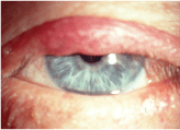
Hordeola and chalazia may result from prolonged meibomian gland disease. Courtesy: Alan G. Kabat, O.D.
In a survey conducted by the National Rosacea Society, 95% of 1,780 rosacea patients with ocular symptoms said that their eyes felt dry, gritty or irritated. Of these, only 28% reported a formal diagnosis of ocular rosacea.1
Meibomitis refers to an inflammation of the meibomian orifices. When severe, it presents as a thick, viscous plug of material. It is not uncommon for patients with ocular rosacea to present with both blepharitis and MGD. This may lead to a hordeolum, or a local infection of meibomian glands or the glands of Zeiss and Moll. Chalazia also frequently occur in such patients.
The conjunctivitis that may occur in ocular rosacea is usually chronic, bilateral and bacterial in nature.8 There is diffuse conjuctival hyperemia, which is often accompanied by lid signs (blepharitis and MGD). Either invading microorganisms or ocular tissues may secrete pseudomembrane consisting of fibrinous inflammatory exudates. The membrane permeates the superficial layers of conjunctival epithelium.
In ocular rosacea, the cornea may be involved to varying degrees. Early findings may include punctate staining (punctate epithelial keratopathy) of the inferior one-third of the cornea. Moderate keratitis is characterized by increased staining and a sterile marginal infiltrate.2
In especially severe cases, advanced keratopathy may ensue with corneal neovascularization, opacification, thinning, ulceration and perforation.
Rosacea Management
Treatment of rosacea depends on both the subtype(s) and present stage/severity of the condition. Tetracycline is often used to treat rosacea. It decreases bacterial lipase, which improves the solubility of sebaceous gland secretions.9
A typical dosing regimen is 100mg doxycycline b.i.d. for six weeks, followed by q.d. for two to four weeks. When longer-term therapy is needed, prescribe 20mg Periostat (doxycycline hyclate, CollaGenex Pharmaceuticals) either q.d. or b.i.d. Periostat was initially developed for periodontitis and is now available in generic form.
Oracea (doxycycline monohydrate, CollaGenex Pharmaceuticals) was FDA approved in April 2006 to treat inflammatory rosacea in adults. Oracea is the first drug to be approved for papulopustular rosacea only. It contains 30mg of immediate-release medication and 10mg of delayed-release medication in one capsule. Oracea exhibits anti-inflammatory, but not antimicrobial, properties, so there are essentially no drug resistance issues.
Tetracycline is contraindicated for children, pregnant women and nursing mothers, and individuals with poor kidney function. Side effects of tetracycline may include upset stomach, photosensitivity and even pseudotumor cerebri.10 Simultaneous ingestion of antacids and dairy products reduces the effectiveness of tetracycline. Erythromycin may be a viable alternative for those cases in which tetracycline is contraindicated or has produced the aforementioned side effects.
Metronidazole is another established medical therapy for rosacea. It exhibits antimicrobial, anti-inflammatory and immunosuppressive properties.11 It is available in oral and topical forms. The drug is effective for inflammatory lesions, not telangiectasias.
Various Ocular Rosacea Treatments Multiple treatments are key: Lid hygiene: Pre-packaged pad scrubs and foams. Hot compresses. Saline soaks. Lubrication with tear supplements: Topical light steroids. Antibiotic/steroid combination (moderate/severe presentation may require a topical fluorquinolone). Restasis (cyclosporine A, Allergan). Doxycycline. Comanagement with primary-care physician. Dermatology consultation for systemic management and laser treatment.
A low-potency topical corticosteroid lotion, such as desonide, may prove useful in certain patients. Retinoids, such as isotretinoin, are vitamin A derivatives that suppress sebum production. This class of drugs has been used for severe or recurrent rosacea. H-2 antagonists that combat Helicobacter pylori have also been implemented.
One drop q.i.d. to q2h.
Surgical treatment for severe corneal complications.
Surgical therapy of rosacea is gaining popularity within the dermatology community. A pulsed-dye laser for erythematotelangiectatic rosacea may be effective in reducing redness and telangiectasia. Surgical ablation, electrocautery and CO2 laser (to remove hypertrophied tissue and reshape the nose) therapy are also available.
Dry Eye and Rosacea
Dry eye associated with rosacea results from evaporative loss due to meibomian gland dysfunction. Researchers have found increased interleukin-1 (IL-1) alpha concentrations in the tears of rosacea patients, as well as greater matrix metalloproteinase (MPP) activity.12 Tetracycline has an inhibitory effect on both these factors.12
So, you may ask: Is there one overarching strategy to address dry eye in rosacea patients? Not really. However, here are some general guidelines to follow:
First, you must address both blepharitis and meibomitis if either condition is present. You can implement supportive therapies for mild signs and symptoms of dry eye. These may include the use of a humidifier at home or in the work environment and tear supplements as often as q2h. Also, implement nutritional support with omega-3 fatty acids, flaxseed oil and increased water intake.
For signs and symptoms of moderate dry eye in rosacea patients, you may want to add topical anti-inflammatory therapy, especially if there is minimal or no improvement with supportive treatments.
A two-week trial can be initiated with light topical steroids, such as Flarex (fluorometholone acetate, Alcon), Lotemax (loteprednol etabonate, Bausch & Lomb) or Pred-Mild (prednisolone acetate 0.12%, Allergan) before starting the patient on long-term treatment with Restasis (cyclosporine A, Allergan).
If topical anti-inflammatory agents result in minimal or no improvement, oral tetracyclines may be considered. The dosage and duration of systemic treatment for ocular rosacea is similar to that for the other sub-types (100mg doxycycline b.i.d. for six weeks). After several weeks of systemic therapy, the dosage may be tapered or the patient should be switched to Oracea.
Lacrimal/punctal occlusion may be implemented after the inflammation is controlled. This can be accomplished with either punctal plugs or cautery. If the patient demonstrates no improvement, minimal improvement, or severe symptoms and signs of ocular rosacea (e.g., severe PEK, corneal erosions or conjunctival scarring), it would be prudent to consult a corneal specialist. Treatment with oral tetracycline and/or cyclosporine may be indicated at this time. A partial or complete tarsorrhaphy may help re-establish optimal tear osmolarity.
Guiding the Patient
As primary eye and health-care providers, optometrists must also educate and, in some cases, counsel their rosacea patients. Instruct the patient to avoid trigger foods such as chocolate, tomatoes and citrus fruits, hot spices, alcohol and heated beverages. Also, tell him or her to avoid direct exposure to sunlight and to use appropriate amounts of sunscreen.
Because of the potential for changes in facial appearance, some patients experience anxiety, depression and poor self-esteem. These people need reassurance and perhaps psychological counseling and/or other therapy from appropriate providers. Stress management may also be helpful.
Ocular rosacea is a distinct condition, with surface inflammatory disease as its most common clinical feature. One of our challenges as clinicians is to effectively diagnose and manage/comanage acute and chronic signs and symptoms. We must communicate and coordinate care with the appropriate physician (either primary care doctor or dermatologist) in a timely and effective manner, which will result in improved patient outcomes.
Dr. Pizzimenti is an associate professor at Nova Southeastern University College of Optometry. Dr. Pelino is an assistant professor at Pennsylvania College of Optometry. Both doctors lecture extensively on oculosystemic disease.
1. National Rosacea Society. Information for Physicians: Patient Education Materials. Available at: http://www.rosacea.org (Accessed May 16 2008).
2. Akpek EK, Merchant A, Pinar V, Foster CS. Ocular rosacea: patient characteristics and
follow-up. Ophthalmology Nov 1997;104(11):1863-7.
3. Murphy G. Histology of the skin. In: Elenitsas R, Jaworsky C, Johnson B, eds. Lever"s Histopathology of the Skin.
4. Stevens G, Lemp M. Acne rosacea. In: Weingeist T, Gould D., eds. The Eye in Systemic
Disease.
5. Wilkin J. Standard Classification of Rosacea. J Am Acad Dermatol 2002 Apr;46(4):584-7.
6. Knox C. Rosacea: A Review of a Common Disorder. The Internet Journal of Academic
Physician Assistants, 2006;4(2).
7. Dahl M, Ross A, Schlievert P. Temperature regulates bacterial protein production: possible role in rosacea. J Am Acad Dermatol 2004 Feb;50(2):266-72.
8. Browning DJ, Proia AD. Ocular rosacea. Surv Ophthalmol 1986 Nov-Dec;31(3):145-58.
9. Nazir S, Murphy S, Siatkowski R, et al. Ocular rosacea in childhood. Am J Ophthalmol. 2004 Jan;137(1):138-44.
10. Frucht-Pery J, Sagi E, Hemo I, Ever-Hadani P. Efficacy of doxycycline and tetracycline in ocular rosacea. Am J Ophthalmol. Jul 15 1993;116(1):88-92.
11. Barnhorst DA Jr, Foster JA, Chern KC, Meisler DM. The efficacy of topical metronidazole
in the treatment of ocular rosacea. Ophthalmology Nov 1996;103(11):1880-3.
12. Afonso AA, Sobrin L, Monroy DC, et al. Tear fluid gelatinase B activity correlates with IL-1alpha concentration and fluorescein clearance in ocular rosacea. Invest Ophthalmol Vis Sci 1999 Oct;40(11):2506-12.
