 Although it may seem unthinkable to many of us in healthcare, patients sometimes seek medical advice from online communities, such as Facebook or Yahoo!, before contacting their doctors.
Recently, the following post came to our attention: “Help! What do you do for a bee sting in the eye? I was just now mowing the lawn when I was dive-bombed by a bee that stung me right on the upper eyelid.”
This month’s column deals with the unusual, but very real, dilemma of managing ocular sting injuries from bees, wasps and other flying insects.
Sting’s Greatest Hits
Sting injuries have long been recognized as a significant source of ocular trauma.1 Bee and wasp stings can directly initiate a variety of ocular manifestations, as well as yield secondary systemic repercussions. In general, injuries from bee stings may be categorized as mechanical, toxic and/or immunologic.2
• Mechanical. The aculeus or “stinger” of bees and wasps (order: Hymenoptera) is a modified ovipositor. Therefore, only female insects are capable of producing sting injuries.3
The stinger is composed of chitin—a tough, keratin-like substance that is the principle component of arthropod exoskeletons. The honeybee’s aculeus is barbed and cannot be withdrawn after an attack. Thus, this structure (and the associated venom sac) is forfeited, resulting in the insect’s death.
While wasps, hornets and bumblebees do not possess a barbed stinger, portions of this structure may occasionally break off and remain in the tissue. So, sting injuries to the ocular structures often result in a puncture wound and a foreign body insult.
• Toxic. The aculeus is attached to a venom sac that is located at the base of the insect’s abdomen. In the act of stinging, a highly toxic, species-specific venom is injected and actively pumped into the tissue of the adversary. The venom of stinging insects is composed primarily of biogenic amines, polypeptides and enzymes.2-4
Envenomation has several effects. Amines (e.g., histamine and dopamine) cause an immediate pain response, presumably to deter further invasion or insult to the insects’ territory.5
Major peptides, such as apamin, bradykinin and melittin, cause cellular damage and disruption of normal metabolic processes, and can serve as potent neurotoxins.2,3 Enzymes, including phospholipase A, hyaluronidase and phosphodiesterase, further exacerbate local damage by perpetuating the immune response.5
• Immunologic. The mechanical trauma and toxic effects of insect venom combine to initiate an inflammatory cascade in the victim of a sting injury. Components of the venom induce an acute response at the inoculation site that involves vasodilation, tissue edema and leukocyte chemotaxis.2,4 With prolonged exposure to the retained stinger, chronic inflammation—as well as secondary local tissue degradation—may ensue.
Systemic allergic reactions can also be seen in hypersensitive individuals, although the prevalence of these allergies is actually quite low. According to the literature, only about 1% to 9% of adults experience systemic reactions to bee stings; children are even less likely to manifest these responses.6,7 Anaphylaxis due to insect venom is exceedingly rare, occurring in less than one in 15,000,000 cases annually.7
Ocular Impact of Stings

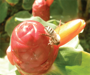
Honeybees can pose a significant risk to ocular health in cases of sting injuries to the eye or adnexa. (This photograph was snapped by Dr. Kabat at Review of Optometry’s annual continuing education meeting in Maui!)
Sting injuries can affect a variety of ocular structures. The lids and adnexa are most commonly involved, and patients typically present with an acutely painful, swollen eye. Redness, edema and lacrimation are the hallmarks of this type of injury. The area may be indurated (e.g., firm and nodular) around the site of inoculation, and the retained stinger may be visible.
Direct injuries to the lid may involve penetration of the tarsus. In such cases, not only is a venomous reaction possible, but also the disembodied stinger may damage the corneal surface.8
Direct stings to the ocular surface, including the cornea or conjunctiva, occur less commonly, but are far more serious. Conjunctival manifestations include acute pain and hyperemia, as well as pronounced edema that is often referred to as “watchglass chemosis.” Retained stingers may represent an additional source of irritation and insult, because they can continue to diffuse venom into the tissue long after the initial traumatic event.
When the cornea is directly involved, patients experience a greater degree of pain and a focal inflammatory reaction will be noted at the site of inoculation. Initially, this may be seen as localized, disciform edema, but tissue degradation will occur as the toxins impact the cellular structure. White cell infiltration is often noted within an hour of the injury, and an anterior chamber response is also common.2
Biomicroscopically, a corneal sting injury may appear similar to a foreign body with infiltrate or even an infectious keratitis.9,10 Deeper repercussions also can be seen. In fact, if the stinger penetrates the cornea, a more severe, toxic uveitis can result.
Even more alarming, several cases of cataract formation secondary to wasp sting penetration through the anterior lens capsule have been documented.1 Cataracts can also develop subsequently as sequelae of chronic, toxicity-induced inflammation.
Other long-term changes include: bullous keratopathy; corneal neovascularization and opacification; persistent uveitis; iris atrophy; and secondary glaucoma.3 In rare instances, involvement of the posterior segment may also ensue. Numerous cases of sting-induced optic neuritis have been reported, as well as retinal damage and ciliochoroidal detachment.4,11-15
Management Strategies
Management strategies for bee and wasp stings vary based upon the location and extent of the injury. For superficial wounds, first aid may be all that is required.
Retained stingers in the skin of the lids or adnexa should be removed carefully and the area should be cleaned with soap and water or Betadine (povidone-iodine, Alcon). Cold compresses may be helpful in limiting the secondary inflammatory response. Ice (wrapped in cloth to avoid freezing the skin) should be applied for no more than 20 minutes every hour.16 Oral antihistamines (i.e., 10mg loratadine) and anti-inflammatory agents (i.e., 200mg to 400mg ibuprofen q.i.d.) may help control associated itching and pain.
Sting injuries involving the globe are more serious and warrant additional therapy. Removal of the stinger with a jeweler’s forceps under biomicroscopy is paramount.
If the conjunctiva is the inoculation site, medical treatment is limited to the ocular surface. Topical antibiotics and corticosteroids—either separate or in combination—represent the ideal therapy for preventing secondary bacterial infection and curtailing associated inflammation.2
Be sure to treat corneal injuries in a similar fashion. Broad-spectrum antibacterial agents, such as gentamicin or a fluoroquinolone, have been advocated.10
In addition, more frequent administration of potent steroids (e.g., prednisolone acetate 1% or difluprednate 0.05%) should be employed to address any concurrent uveitis. Cycloplegia with a strong agent, such as scopolamine or atropine, is likewise recommended. Treating uveitis aggressively and proactively will help prevent long-term sequelae, including cataracts, iris atrophy and glaucoma.
For patients with toxic optic neuritis associated with envenomation, early intervention with pulsed steroids may prevent permanent visual loss.10-13
A three-day course of intravenous methylprednisone followed by a tapered dose of oral prednisone over seven additional days has been shown to help patients recover visual function following this type of trauma.12
While ocular sting injuries are not a common occurrence, primary eye care providers must be ready for any scenario. Recognition, careful evaluation and prompt intervention in these cases can help prevent or reverse significant visual impairment.
Drs. Kabat and Sowka are paid consultants for Alcon Laboratories. They have no direct financial interest in any of the products mentioned.
1. Gilboa M, Gdal-On M, Zonis S. Bee and wasp stings of the eye. Retained intralenticular wasp sting: A case report. Br J Ophthalmol. 1977 Oct;61(10):662-4.
2. Lin PH, Wang NK, Hwang YS, et al. Bee sting of the cornea and conjunctiva: management and outcomes. Cornea. 2011 Apr;30(4):392-4.
3. Arcieri ES, França ET, de Oliveria HB, et al. Ocular lesions arising after stings by hymenopteran insects. Cornea. 2002 Mar;21(3):328-30.
4. Choi MY, Cho SH. Optic neuritis after bee sting. Korean J Ophthalmol. 2000 Jun;14(1):49-52.
5. Valentine MD. Insect venom allergy: diagnosis and treatment. J Allergy Clin Immunol. 1984 Mar;73(3):299-304.
6. Antonicelli L, Bilò MB, Bonifazi F. Epidemiology of Hymenoptera allergy. Curr Opin Allergy Clin Immunol. 2002 Aug;2(4):341-6.
7. Bilò MB. Anaphylaxis caused by Hymenoptera stings: from epidemiology to treatment. Allergy. 2011 Jul;66 Suppl 95:35-7.
8. Chaurasia S, Muralidhar R. Retained bee stinger in the tarsal plate. Int Ophthalmol. 2011 Apr;31(2):111-2.
9. Jain V, Shome D, Natarajan S. Corneal bee sting misdiagnosed as viral keratitis. Cornea. 2007 Dec;26(10):1277-8.
10. Chinwattanakul S, Prabhasawat P, Kongsap P. Corneal injury by bee sting with retained stinger--a case report. J Med Assoc Thai. 2006 Oct;89(10):1766-9.
11. Berríos RR, Serrano LA. Bilateral optic neuritis after a bee sting. Am J Ophthalmol. 1994 May 15;117(5):677-8.
12. Maltzman JS, Lee AG, Miller NR. Optic neuropathy occurring after bee and wasp sting. Ophthalmology. 2000 Jan;107(1):193-5.
13. Kim JM, Kang SJ, Kim MK, et al. Corneal wasp sting accompanied by optic neuropathy and retinopathy. Jpn J Ophthalmol. 2011 Mar;55(2):165-7.
14. Kitagawa K, Hayasaka S, Setogawa T. Wasp sting-induced retinal damage. Ann Ophthalmol. 1993 Apr;25(4):157-8.
15. Pal N, Azad RV, Sharma YR, et al. Bee sting-induced ciliochoroidal detachment. Eye (Lond). 2005 Sep;19(9):1025-6.
16. WebMD. Treatment of Bee and Wasp Stings: First Aid & Emergencies. Available at:
http://firstaid.webmd.com/bee-and-wasp-stings-treatment (accessed August 28, 2011).

