 |
An 89-year-old white female was referred by her primary care physician for evaluation of an acute herpes zoster infection with diplopia and ptosis. She reported that a rash developed around her left eye and forehead 10 days prior, and that her left eyelid started to droop three days prior. She would experience double vision when she lifted her eyelid. She also reported left eye pain, blurry vision and significant neuralgia around the left side of her head. Her primary doctor started her on Valtrex (valacyclovir hydrochloride, GlaxoSmithKline) and gabapentin. Her medical history was remarkable for hypertension, for which she was taking triamterene/hydrochlorothiazide. Her ocular history included left central retinal vein occlusion since 2013 with chronic macular edema, treated with Ozurdex (dexamethasone intravitreal implant, Allergan) and Eylea (aflibercept, Regeneron) injections.
Her best-corrected visual acuities were 20/25 OD and 20/100 OS. Intraocular pressure (IOP) was 18mm Hg in each eye. Her pupils were equal, round and reactive to light with no afferent pupillary defect noted. A resolving vesicular rash and erythema was noted along the left ophthalmic branch of the trigeminal nerve. A complete left ptosis was noted. Extraocular motilities showed restricted upgaze, downgaze and adduction of the left eye.
A slit lamp examination of the right eye was unremarkable. The left eye showed 1+ conjunctival injection and ciliary flush with a 1+ anterior uveitis. Her cornea was clear and her posterior segment was quiet. The retinal findings in the left eye were consistent with her report of an old retinal vein occlusion. OCT showed no macular edema.
Diagnosis & Management
We diagnosed our patient with a complete, pupil-sparing left 3rd nerve palsy secondary to herpes zoster. Antivirals are the mainstay therapy for herpes zoster infections. We typically prefer either valacyclovir or famciclovir over acyclovir because they are more effective in limiting postherpetic neuralgia.1 She was already taking Valtrex 1g TID per her internist, so we continued her on the current dose. Additionally, because the suggested mechanism for zoster-related oculomotor nerve palsies is inflammatory in nature, we started her on prednisone 60mg QD and treated her uveitis with Durezol (difluprednate ophthalmic emulsion 0.05%, Alcon) QID OS.
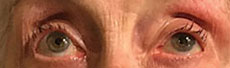
Two weeks later, she showed a 60% to 70% improvement in her ptosis and an 80% to 90% improvement in the extraocular movements. Her ocular inflammation was also completely resolved and vision improved to 20/30 in the left eye. We cut the Valtrex to 500mg TID for an additional 10 days and started a prednisone and Durezol taper. Three weeks later, the 3rd nerve palsy had completely resolved with no recurrence. Even with very long tapering of the steroid drops, our patient developed a chronic recurring herpetic uveitis. Because of our concerns for complications, especially with her history of chronic macular edema from a vein occlusion, we decided to treat her with prednisolone acetate QD, which has kept the eye quiet. Her vein occlusion and macular edema have remained stable.
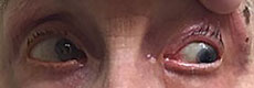
Initial Visit and Follow-upInitial visit
At initial visit, ptosis with restricted upgaze, downgaze and adduction. Follow-up
|


Discussion
When herpes zoster infection affects the ophthalmic branch of the trigeminal nerve, it is referred to as herpes zoster ophthalmicus (HZO).2 Ocular complications of HZO occur in approximately 50% of cases and commonly include keratitis, pseudodendrite, episcleritis, uveitis and increased IOP.2,3 Although uncommon, cranial nerve palsies have been seen in patients with HZO. Cranial nerves 3, 4 and 6 have all been reported but cranial nerve 3 is most commonly affected—involved in 47% of all cases.2,3 On average, the nerve palsy occurs nine and a half days after the onset of rash with a range of two to 42 days.2 Although the exact mechanism is unclear, research suggests inflammation of the trigeminal nerve could spread to the other cranial nerves within the cavernous sinus.4 Research also suggests occlusive vasculitis as a culprit.5
Though not seen in our patient, oculomotor nerve palsies associated with HZO can affect the pupil. If an aneurysm or space-occupying lesion is suspected, especially with pupillary involvement, MRI and MRA are suggested. Our patient’s age, lack of pupillary involvement, and the palsy’s timing in relation to the shingles infection and treatment response made aneurysm unlikely in our estimation; therefore, neuroimaging was not performed.
HZO can present with a variety of ocular complications and can be challenging to manage at times. Understanding the various complications and instituting the proper treatment will help improve patient outcomes.
|
1. Pavan-Langston D. Herpes zoster antivirals and pain management. Ophthalmol. 2008 Feb;115(2 Suppl):13-20. 2. Harthan J, Borgman C. Herpes zoster ophthalmicus-induced oculomotor nerve palsy. J Optom. 2013 Jan;6(1):60-5. 3. Hakim W, Sherman R, Rezk T, Pannu K. An acute case of herpes zoster ophthalmicus with ophthalmoplegia. Ophthalmol Med. Epub 2012 May 9. 4. Edgerton A. Herpes zoster ophthalmicus: report of cases and a review of the literature. Transactions of the Amer Ophthalmol Soc. 1942;40:390-439. 5. Naumann G, Gass M, Font R. Histopathology of herpes zoster ophthalmicus. Am J Ophthalmol. 1968;65(4):726-9. |
