Your patients’ food choices and dieting patterns can sometimes be overwhelming. Today, new dietary trends, such as paleo or gluten-free, and specific labeling, such as hormone-free and free range, can be confusing to patients simply looking for the healthiest diet—and to practitioners managing patient health. By understanding the nutritional makeup of food and its impact on systemic and ocular health, practitioners can better educate patients on their dietary choices and how to protect their ocular health.
Here is a look at what the body needs, nutritionally, and what many of today’s common diets do to the body—and the eye.
 |
| Patients need to maintain the proper levels of macronutrients, vitamins and minerals through their diets. |
Dietary Basics
Most diets can be healthy, as long as they are done properly by ensuring sufficient intake of necessary vitamins and nutrients in meals or, if needed, supplements. The three main components of a proper diet are: protein, carbohydrates and good fats. Proteins help the body maintain muscle mass, speed up metabolism and feel full longer. Carbs provide quick energy when the body breaks them down into single glucose molecules, which are then stored for later use. Lastly, fats keep you warm and provide you with essential fatty acids, which help to control inflammation and aid in blood clotting and brain development.1 Fats also help the body absorb fat-soluble vitamins A, D, E and K.1
Vitamins and minerals. The United States government provides recommended daily values for all vitamins and nutrients. Each one has a job in helping our bodies and eyes grow and function
properly.
From recent studies such as the AREDS I and II, we know that zeaxanthin and lutein help to reduce chronic eye disease, filter harmful blue light and act as natural macular antioxidants to help decrease progression of severe age-related macular degeneration (AMD).2 The body uses alpha lipoic acid to prevent certain types of cell damage, restore vitamin levels and break down carbohydrates to improve insulin sensitivity.3 Zinc is important for fetal development, wound healing, DNA synthesis and the immune system.3 It is found in high amounts within the retina, and studies show zinc deficiency causes dark adaptation defects.4,5 Vitamin A, which is critical to the immune system’s function, also aids in retinal melanin production.6 Deficiency can lead to night blindness, which is often accompanied by Bitot’s spots on the conjunctiva.6 In severe forms of deficiency, corneal ulceration can occur along with keratomalacia.6,7,3
Vitamin C is associated with a lower risk of cataract formation by protecting the proteins of the crystalline lens from oxidation.8 Research also suggests it acts as an antioxidant, inhibiting retinal cell damage from free radicals liberated by UV exposure.5 It also helps to develop connective tissue and biosynthesize collagen, both of which aid corneal wound healing.8 Vitamin C keeps neurotransmitters functioning and, of course, boosts immune function.8 Vitamin E also helps with immune function and protects cells from free radicals by acting as an antioxidant, which helps to delay or prevent chronic systemic and retinal diseases.5,8 Deficiency of this vitamin has been identified as part of the pathogenesis of AMD, though supplementation of vitamin E alone was not found to decrease incidence.2,5,8
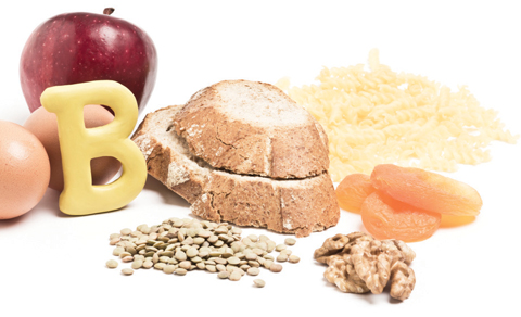 |
| B vitamins play a large role in maintaining ocular and systemic health. |
As with other vitamins and minerals, vitamin K bears clinical significance, as blood clotting factors are affected by vitamin K absorption, which can cause concern for retinal bleeding.3
Several B vitamins exist, and each plays an important role in the body and the eye:
• B1 (thiamine) helps in energy metabolism but also with the function of the central nervous system.3 Deficiency can lead to Wernicke’s encephalopathy with nystagmus, rectus muscle fatigue or paralysis, optic atrophy and loss of visual acuity.3,9
• B2 (riboflavin) also aids in metabolism by facilitating amino acid production and acting as a cofactor in antioxidant production.3 Lack of riboflavin can lead to corneal epithelial dystrophy, vascularization of the cornea, angular blepharoconjunctivitis and decreased vision.3,9
• B6 (pyridoxine) influences red blood cell production, brain function, immune function and decreased homocysteine.3,9 A deficiency in this vitamin can lead to optic neuritis and angular blepharoconjunctivitis.9
• B7 (biotin) helps to mediate cell growth, fatty acid production, fat metabolism and steady blood sugar levels.3,9 This is found in concentrated quantities within the retina, aiding in the maintenance of retinal health.5
• B9 (folic acid), critical for fetal development, also combats anemia and aids in red blood cell production.3 Low levels can lead to nutritional optic neuropathy.5,9
• B12 (cobalamin) is used for the essential task of myelin synthesis and repair and DNA/RNA production.3 Research shows some diabetes patients taking metformin are at increased risk of B12 deficiency as a side effect of the medication.10,11 Studies show abnormal B12 metabolism in patients with peripheral neuropathy, diabetic retinopathy and optic neuropathy.4,9,10,11
Essential fatty acids (EFAs). Multiple studies, including the Dream Study, show the importance of essential fatty acids in treatment of dry eye disease (DED), which inhibit T-lymphocyte production associated with the disease and pro-inflammatory cytokines, IL-1, IL-2 and TNF-alpha.12,13,14 EFAs such as omega-3s, which are components of cell membranes, aid visual development and maintain retinal function, increase meibomian gland secretions, boost the immune system, decrease inflammation, maintain the nervous system’s integrity and decrease cholesterol levels.2,12,14-17 One study observed a correlation between low dietary intake of omega-3 EFAs and DED in women, and a 30% decrease in DED with each additional gram of omega-3 EFAs consumed daily.18
 |
| The popular paleo diet may alleviate a common issue with the American diet: pre-diabetes and diabetes. However, this dietary trend isn’t without its risks. |
The Risks
Popular diets as well as dietary lifestyles come with both risks and benefits. Here’s a breakdown of the most popular diets and dietary habits, how much (or little) of their popularity is based on myth or fact, and how to best make dietary changes without hurting ocular health:
Paleo. This diet’s rather simplistic mantra is, “if a caveman couldn’t eat it, neither can you.”18,19 Thus, all processed foods are avoided, including grains, legumes, added salt, dairy and alcohol. Although the diet hasn’t been around long, the idea of eating clean unprocessed foods is not new. The mainstays of the diet include fresh fruits and vegetables, meats, seafood and nuts. According to one study, the right balance should be approximately 35% of calories coming from fat, 35% from carbohydrates and 30% from protein.21 Maintaining this diet may help the body decrease its glycemic load through a healthy ratio of saturated to unsaturated fatty acids, an increase in vitamin and nutrient consumption, and an optimal balance of protein, fat and carbs.20 Other studies have found the paleo diet improved blood sugar control over 12 weeks compared with a Mediterranean diet, and overall better glucose tolerance; long-term, this may reduce risk for type 2 diabetes.19,20
Concerns with the diet include the risk of consuming too much protein; a 1:1 ratio of calories from meat to that of produce can lead to kidney damage.22 Consuming too much saturated fat from red meat can also lead to heart disease.23 Since dairy is not a part of paleo, patients on the diet could be low on calcium and vitamin D, which can lead a higher risk for osteoporosis, hypertension and diabetes.24 Some research shows a lower risk of MS in Caucasians with high levels of vitamin D, with increasing incidence rates in correlation to distance from the equator. Therefore, researchers hypothesize that vitamin D may mediate an anti-inflammatory immune response, specifically enhanced regulatory T-cell function.24
Ocular involvement: Research shows a high amount of meat protein, a common concern with paleo, is linked to a higher risk of early cataract development.25 Vitamin D deficiencies associated with this diet may also lead to an increased risk for of AMD, which is thought to result from the increased inflammation and angiogenic effects that occur in the absence of the vitamin.26 These then contribute to early retinal vascular damage.26 One study also suggests the impaired immunity caused by vitamin D deficiency increases risks for corneal diseases such as herpes simplex zoster and contact lens-associated acanthomoeba.24,26
Vegetarian/vegan. While vegetarian diets can vary significantly, their defining characteristic is abstention from the consumption of meat, including red meat, poultry, seafood and flesh of any animal. Ovo-lacto vegetarians eat eggs and dairy, ovo- vegetarians eat eggs but no dairy and lacto-vegetarians eat dairy but no eggs. Vegans abstain from eating animals or any animal byproducts (as well as refraining from use of any products made from animals, including wool and leather).
Vegetarian and vegan diets, when done properly, tend to be lower in fat and cholesterol with higher levels of fiber and antioxidants. This has been associated with a lower body mass, lower rate of death from heart disease and lower levels of cholesterol.27 Research also suggests there is a lower rate of hypertension, type 2 diabetes mellitus and prostate and colon cancer in vegetarians.27
Ocular involvement: In a 2011 study, researchers found a correlation between a lower risk of cataract development and a lower consumption of meat.28 However, as reported by the Academy of Nutrition and Dietetics, the lifestyle is, at times, also associated with low dietary levels of protein, iron, zinc, calcium, vitamin D, riboflavin, vitamin B12, vitamin A, omega-3 and iodine.9 Such shortages may contribute to ocular issues; night blindness or corneal ulcerations arise from vitamin A deficiency, low EFA intake worsens DED and MGD and optic neuropathy can arise with vitamin B12 deficiencies, for example.7,16,17,28
Gluten-free. While some patients eliminate gluten from their diet due to rumors that it’s the root of all modern maladies, in several cases, valid medical reasons exist for avoiding gluten: wheat allergy, non-celiac gluten sensitivity (NCGS) and celiac disease. Wheat allergy is an immune reaction to a wheat protein, mediated by immunoglobulin E (IgE) antibodies that are sent out to attack the wheat.29 NCGS, as its name suggests, is a sensitivity to gluten, which causes intestinal and extraintestinal distress that is not IgE mediated or an autoimmune reaction.30-32 It is currently a diagnosis made by exclusion.31,32
Celiac disease is an autoimmune disorder triggered by ingestion of gluten. Affected individuals must inherit the genetic predisposition, be consuming gluten and have the disease activated. Activation usually occurs secondary to stress, trauma or surgery, and sometimes viral infections.33-35 When a person with activated celiac consumes gluten, the cells lining the small intestine mal-absorb nutrients, which may cause deficiencies.
Ocular involvement: Wheat is sometimes found in cosmetics and lotions, causing a wheat allergy to manifest as allergic conjunctivitis, eye irritation and eczema both of the lids and the periorbital skin.36,37 Most commonly, malabsorption associated with celiac disease occurs with vitamin A, vitamin D and calcium, leading to retinopathy, night blindness, an increase in cataracts, DED and, occasionally, idiopathic intracranial hypertension.8 The autoimmune disorder-related ocular effects include orbital myositis, uveitis, thyroiditis with orbitopathy and brain occipital calcifications.33 While ocular issues associated with the disease itself may not be avoidable, making sure patients increase their dietary or supplementary vitamins A, D, B and calcium can help them avoid some of the other gluten-free complications.
 |
| Wheat, a staple of the American diet, is intolerable to some due to gluten allergy, non-autoimmune gluten sensitivity and celiac disease. |
Health Mantra
When counseling a patient regarding a specialty diet, as with all things, the key word is moderation. The mantra for any of your patients on a special diet should be: read your labels and supplement any missing vital vitamins and nutrients.
Labels. Patients have to wade through many new food labels with a wide range of implications. Here is a quick look at what some of these new labels actually mean.
Cage-free indicates that the animal has never been contained in a cage, while free range animals have open access to food and water, with allowance to roam in certain defined spaces.38
USDA-labeled organic foods have a checklist of criteria that differentiate them from natural, including:1
• no toxic persistent pesticides
• no genetically modified organisms (GMOs) in the feed
• no antibiotics
• no sludge or irradiation
• meets specific animal welfare requirements (e.g., cows are required to be on pasture for pasture season)
• contains low levels of environmental pollution
All of these are closely monitored with audit trails as well as inspection certifications.
Hormone-free and claims of no antibiotic use are voluntary FDA labeling commonly used with milk and other dairy products to indicate that the cows were not treated with growth hormones. Three levels within these categories exist: (1) raised without antibiotics, (2) not fed antibiotics and (3) no detectable antibiotic residue.
GMO is a voluntary FDA label that indicates the organism (plant, animal or microorganism) has had genetic material altered in a way that did not happen naturally through mating or natural recombination. Studies show these foods can contain increased amounts of dietary vitamin A and carotenoids, which decrease the risk of certain diseases, including retinal degenerations.39
 |
| Though chewable supplements improve compliance, studies show they contain lower levels of vitamins and minerals. |
Supplementation shortcomings. The body always absorbs nutrients and vitamins more readily from food, so the best source is the diet. Foods lose nutrient and vitamin content during processing, storage and milling, so fresh is better.40 While a deficiency of vitamins and minerals exposes the individual to potential systemic and ocular problems, the opposite can be true as well. For instance, excessive amounts of vitamin A can cause increased cerebrospinal fluid pressure, papilledema, double vision and dry mucous membranes.1,3,7 While taking excessive amounts of water-soluble nutrients may cause some uncomfortable side effects, such as nausea and diarrhea with vitamin C, they most likely pose little ill effects. On the other hand, fat-soluble vitamins—A,D,E and K—pose real risks for toxicity.41 Therefore, instruct patients to adhere to the proper dosages listed on their supplements, assuming their supplements are up to par with the standards for quality and efficacy.
Additionally, while dietary supplements can help make up for what’s lacking in the diet, the body’s absorption of these items varies based on composition. And, while all supplements contain labels, the actual contents vary.
To ensure patients are getting the best value, clinicians should recommend specific supplements by looking for independent certification labels from the Natural Products Association (NPA), US Pharmacopeial Convention (USP) and NSF. Independent chemical analysis websites such as labdoor.com and consumerlab.com break down the contents of many brands to assess the accuracy of labels, product purity, ingredient safety and potential efficacy. For example, labdoor.com found the average tested values of vitamin label claims were off by 22.6% and minerals by 29.2%. The most overstated amounts were vitamins A and C. Gummy and other chewable multivitamins had 54% less vitamin content and 70% less minerals than their standard
counterparts.42
For EFA supplements in particular, the key is the level of docosahexaenoic acid (DHA) and its precursor, eicosapentaenoic (EPA). AREDS2 used the dosage of 350mg DHA and 650mg EPA per day, while the Dream Study used 1,000mg DHA 2000mg EPA per day.14 Gamma-Linolenic acid, a healthy omega-6, shows benefit in dry eye disease, including in Sjögren’s
syndrome.43
Although no consensus on dosage exists, most studies support 1,000mg of DHA and 1,000mg of EPA daily as adequate to support a healthy ocular system.16,17,44
Krill oil is another alternative for omega-3 supplementation. It is more concentrated and a smaller pill size, which is often more palatable for patients. A recent study shows that after three months of supplementation with either krill oil at a dosage of 945mg EPA and 510mg DHA per day or fish oil at 1,000mg EPA and 500mg DHA, study participants had improved tear osmolarity and increased tear stability compared with placebo.45
Popular diets and dietary habits will come and go, but the body’s nutritional requirements will not change. When clinicians look out for patients’ dietary deficiencies and educate on moderation, we help ensure their ocular health.
Dr. Koetting is the referral optometric care & externship program coordinator at Virginia Eye Consultants in Norfolk, VA. She is a fellow of the American Academy of Optometry and a trustee of the Virginia Optometric Association.
1. Combs GF, McClung JP. The vitamins: fundamental aspects in nutrition and health. Westborough, Mass: Academic Press; 2016. |
