 History
A 46-year-old black male presented for an emergency examination following blunt trauma to his face and left eye after falling down some stairs. His chief complaint was pain and poor vision O.S.
His ocular history was significant for a peripheral retinal tear that required repair and a choroidal rupture that resulted in scarring of the macula O.D.
On a separate occasion, the patient reported that he was struck on the left side of his face with a golf club, which caused a traumatic hyphema and lens subluxation O.S.
Following the golf club incident, the patient underwent vitrectomy and lens removal O.S. and received a sutured posterior chamber intraocular lens implant. His general medical history was noncontributory. He takes no medications and has no drug allergies.
Diagnostic Data
The patients best-corrected visual acuity was 20/400 O.D. and hand motion O.S. External examination of the adnexa revealed old scars that surrounded the right orbit and significant ecchymosis and edema of the left eye. A bony deformity was noted on palpation of the superior orbital rim O.S.
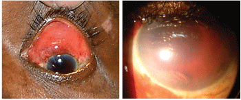
The left eye of our patient who complained of pain and poor vision. Close-up (right).
His intraocular pressure (IOP) measured 18mm Hg O.D. and 0mm Hg O.S. Ocular motilities of the left eye were painful and restricted in all directions of gaze.
The extent of ocular trauma severely limited our ability to observe pupil function, as well as the anterior segment O.S. The patients left anterior segment is pictured above.
Your Diagnosis
How would you approach this case? Does this patient require any additional tests? What is your diagnosis? How would you manage this patient? What is the likely prognosis?
Thanks to Marc Myers, O.D., of
The Diagnosis
The diagnosis in this case is a ruptured globe. The rupture occurred 5mm from the superior limbus and produced a hyphema O.S. We placed a Fox Eye Shield over the left eye and referred the patient to a local tertiary eye care hospital. A computed tomography (CT) scan confirmed an orbital floor fracture. B-scan ultrasonography revealed that the posterior chamber intraocular lens was subluxed.1-4
Acute surgical intervention included repair of the ruptured globe and removal of blood from the anterior chamber O.S. Postoperative medications included atropine 1% b.i.d., prednisolone acetate 1% q2h and ofloxacin 0.3% q2h O.S.1-4
There was grade 2+ corneal edema and an acute postoperative IOP spike of 58mm Hg that warranted the use of oral acetazolamide 500mg b.i.d., brimonidine 0.15% t.i.d. and betaxolol 0.25% b.i.d. O.S. We planned a second surgery to repair the subluxed lens.1-4
During follow-up, we paid special attention to prophylaxis against both infection and corneal edema. We also closely monitored his IOP. The prognosis for this patient in regaining premorbid visual function remains guarded.1-4
1. Hatton MP, Thakker MM, Ray S. Orbital and adnexal trauma associated with open-globe injuries. Ophthal Plast Reconstr Surg 2002 Nov;18(6):458-61.
2. Pump-Schmidt C, Behrens-Baumann W. Changes in the epidemiology of ruptured globe eye injuries due to societal changes. Ophthalmologica 1999;213(6):380-6.
3. Ahmadieh H, Soheilian M, Sajjadi H, et al. Vitrectomy in ocular trauma. Factors influencing final visual outcome. Retina 1993;13(2):107-13.
4. Parnell JR, Small KW. Macular Dystrophies. In: Yanoff M and Duker JS (eds). Ophthalmology.
