 On clinical examination, Salzmann’s nodular degeneration (SND) appears as superficial, bluish-white, raised lesions that are often located in the peripheral cornea. Patients can experience irritation, such as dryness and foreign body sensation, as well as decreased vision in advanced cases.1
A diagnosis of SND carries a very favorable prognosis, and patients usually want to know how the condition can be treated. Here, we’ll review current research about the identification and treatment of SND.
The Specifics of SND
SND is a rare, non-inflammatory, slowly progressive, degenerative condition.2 Women are significantly more likely to experience SND than men.3,4 The average age of presentation is 59 years, and the most common symptom associated with SND is a foreign body sensation.3 Further, the condition presents bilaterally in 63% of cases.4
Confocal microscopy indicates that these lesions are elongated basal epithelial cells and activated keratocytes, particularly in the area of the anterior stroma near the nodules.5 Occasional sub-basal nerves and tortuous stomal nerve bundles are observed. Spectral domain OCT imaging often reveals fibrous intraepitheialial nodules with significant overlying epithelial thinning, which likely contributes to patients’ symptoms.6
Although the patient’s environment may play a role, research suggests that there may be a genetic or familial pattern of development.7
Potential Associations
Recent research showed an extremely high association between patients diagnosed with SND and osteroporosis—suggesting that SND may indicate an abnormality of the patient’s mucopolysaccharides.8 Separate studies have shown a high correlation to chronic ocular surface inflammatory conditions, such as keratconjunctivitis sicca, exposure keratopathy and pterygium.4 Other researchers have questioned if hyaline degeneration of the cornea might be a precursor to SND.9 And yet other studies show a high correlation of SND in patients with epithelial basement membrane dystrophy.10
Topical Treatment Options
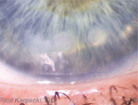
This patient presented with signs of Salzmann’s nodular degeneration.
If patients experience minimal irritation, several topical medications can be helpful. Artificial tears––especially more viscous formulations with longer retention times, such as FreshKote (Focus Laboratories) or Blink Gel Tears (AMO)––may help reduce irritation and discomfort. If patients demonstrate symptoms of grittiness, foreign body sensation or even photophobia, topical corticosteroids (e.g., loteprednol 0.5%) and topical NSAIDs (e.g., bromfenac) can be used to relieve the inflammation and pain.
For long-term management, patients could be treated with cyclosporine A. At this point, topical NSAIDs and corticosteroids would be reserved for more significant symptom episodes.
Surgical Treatment Options
If, however, the nodules are creating significant visual disturbances by severely altering the tear film or causing irregular astigmatism to the cornea, surgery is recommended.
One of the most successful treatments is either superficial keratectomy (SK) or phototherapeutic keratectomy (PTK) with the application of mitomycin C.11 Mitomycin C prevents the formation of corneal haze and/or scarring. In one study, these surgical procedures for SND improved mean best-corrected visual acuity from 20/80 to 20/30.12 Keep in mind, however, that when these peripheral lesions are removed, the patient may potentially experience a myopic refraction shift.13
When a patient presents for surgical removal via SK or PTK, the nodules often separate from Bowman’s layer very easily. Other times––especially when vascularization or pannus is present––the SND lesions can leave a large defect that typically will require excimer laser treatment to remove any irregularity.2 The recurrence rate of SND following PTK is low and estimated to be about 20% within 12 years of surgery.14 Fortunately, corneal and lamellar transplantation is rarely required for patients with SND.15
O.D.s in primary care settings often diagnose SND, so it is imperative to understand its pathogenesis and associated manifestations. Being able to monitor and educate patients, as well as suggest an appropriate treatment, is paramount.
Dr. Karpecki is a paid consultant to Allergan, Bausch + Lomb, AMO, Focus Laboratories and ISTA Pharmaceuticals. He has no direct financial interest in any product mentioned.
1. Lanier JD. Salzmann’s nodular degeneration (subepithelial fibrosis). Ophthalmic Surg Lasers Imaging. 2011 Jul-Aug;42(4):350-2.
2. Das S, Link B, Seitz B. Salzmann’s nodular degeneration of the cornea: a review and case series. Cornea. 2005 Oct;24(7):772-7.
3. Hamada S, Darrad K, McDonnell PJ. Salzmann’s nodular corneal degeneration (SNCD): Clinical findings, risk factors, prognosis and the role of previous contact lens wear. Cont Lens Anterior Eye. 2011 Aug;34(4):173-8.
4. Fario AA, Halperin GI, Sved N, et al. Salzmann’s nodular corneal degeneration clinical characteristics and surgical outcomes. Cornea. 2006 Jan;25(1):11-5.
5. Roszkowska AM, Aragona P, Spinella R, et al. Morphologic and confocal investigation on salzmann nodular degeneration of the cornea. Invest Ophthalmol Vis Sci. 2011 Jul 29;52(8):5910-9.
6. Hurmeric V, Yoo SH, Karp CL, et al. In vivo morphologic characteristics of Salzmann nodular degeneration with ultra-high-resolution optical coherence tomography. Am J Ophthalmol. 2011 Feb;151(2):248-56.e2.
7. Khan K, Ali M, Ingelhearn C. Familial pattern of Salzmann-type nodular corneal degeneration – a four generation series. Reply to Papanikolaou et al. Br J Ophthalmol. 2011 Jun;95(6):890; author reply 890.
8. Nagy M, Hadhazv C, Vigvary L. General symptoms of cornea Salzmann-type degeneration. Orv Hetil. 2007 Mar 4;148(9):413-9.
9. Frising M, Piz S, Olbert D, et al. Is hyaline degeneration of the cornea a precursor of Salzmann’s corneal degeneration? Br J Ophthalmol. 2003 Jul;87(7):922-3.
10. Werner LP, Issid K, Werner LP, et al. Salzmann’s corneal degeneration associated with epithelial basement membrane dystrophy. Cornea. 2000 Jan;19(1):121-3.
11. Koch DD. J Cataract Refract Surg. Impact of Salzmann’s lesions on corneal curvature. 1995 Mar;21(2):111-2.
12. Kharieddin R, Katz T, Baile RB, et al. Superficial keratectomy, PTK, and mitomycin C as a combined treatment option for Salzmann’s nodular degeneration: a follow-up of eight eyes. Graefes Arch Clin Exp Ophthalmol. 2011 Aug;249(8):1211-5.
13. Rimmer S, Maloney RK. Myopic shift after removal of Salzmann’s nodular degeneration. J Cataract Refract Surg. 1994 Nov;20(6):656-7.
14. Das S, Langenbucher A, Pogoreloy P, et al. Long-term outcome of excimer laser phototherapeutic keratectomy for treatment of Salzmann’s nodular degeneration. J Cataract Refract Surg. 2005 Jul;31(7):1386-91.
15. Sinha R, Chhabra MS, Vajpayee RB. Recurrent Salzmann’s nodular degeneration: report of two cases and review of literature. Indian J Ophthalmol. 2006 Sep;54(3):201-2.
