 A 20-year-old white individual was referred for evaluation and management of reduced visual acuity O.D. The patient reported a slow, painless, progressive reduction in acuity over the past four months.
The patient thought that corrective glasses would yield improved vision; however, the eye doctor said that the problem could not be corrected with glasses.
The patient reported being in excellent health, and took no medications. The patients past ocular history is noncontributory. Also, the patient reported always experiencing good vision, and noted last having an eye exam about 10 years ago.
Diagnostic Data
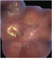
1. A wide-angle fundus montage shows peculiar retinal changes O.D.
On examination, the patients best-corrected visual acuity measured 20/50 O.D. and 20/20 O.S. Extraocular motility testing was normal. Confrontation visual fields were full to careful finger counting O.U. The pupils were equally round and reactive, with no afferent pupillary defect. The anterior segment exam was unremarkable.
A dilated fundus exam of both eyes showed a small cup with good rim coloration and perfusion O.U. Changes were seen in the right eye temporal to the macula (figure 1). There was also an area of interest along the superior temporal arcade.
Additionally, optical coherence tomography (OCT) was performed (figure 2).
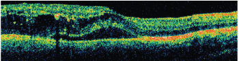
2. A horizontal OCT slice through the macula of the right eye of our patient.
Take the Retina Quiz
1. Based on the clinical presentation, what is the likely gender of our patient?
a. Female.
b. Male.
c. Could be either male or female.
d. Cannot answer based on the information provided.
2. How would you describe the changes that surround the exudate temporally?
a. Retinal neovascularization.
b. Shunt vessels.
c. Macroaneurisms and telangiectasia.
d. Retinal angiomas.
3. What doesnt the OCT show?
a. Retinal thickening.
b. Cystoid macular edema (CME).
c. Serous retinal detachment.
d. Pigment epithelial detachment.
4. What is the correct diagnosis?
a. Old branch retinal vein occlusion (BRVO).
b. von Hippel-Lindau disease.
c. Coats syndrome.
d. Macroarterial aneurysms.
5. How should this patient be treated?
a. Observation.
b. Focal laser photocoagulation.
c. Full panretinal photocoagulation.
d. Pars plana vitrectomy.
For answers, see below.
Discussion
On clinical exam, there is a large circinate ring of exudate that extends into the macula O.D. This exudate is causing retinal thickening, CME, and a serous detachment that involves the macula. Within the area of exudate, there are multiple aneurismal dilations of the retinal vessels and smaller areas of retinal telangiectasia of the smaller retinal vessels. These blood vessel changes are not isolated to this area of the retina. There are similar changes located superiorly above the superotemporal arcade O.D. Interestingly, these changes are isolated only to the right eye.
One of the quiz questions asked about the likely gender of our patient. To be able to answer that question, you need to know the correct diagnosis and what is causing these changes.
In this case, our male patient has Coats disease, which is also known as Coats syndrome. Coats disease is an idiopathic vascular anomaly that is characterized by aneurismal dilations and telangiectasia of the retinal vessels.1,2 It tends to be unilateral and occurs almost exclusively in men (more than 90% of cases).1,2
The retinal capillaries are typically most affected. However, changes can also be seen in the major retinal vessels, particularly the arteries, as is the case in our patient. These vessels become incompetent and leak fluid in the form of exudate. The hallmark of this condition is an exudative retinopathy. The extent of involvement and severity may vary.
In milder forms of Coats disease, only isolated areas of the retina are involved, and the prognosis for maintaining good vision is usually quite good. In more severe cases, larger areas of the retina are involved, and patients can present with sizeable amounts of exudate. Severe Coats disease is more common in children, in which cases most or all the retinal vessels are involved.1 Severe involvement may cause massive subretinal exudate throughout the retina and/or exudative retinal detachments. The visual morbidity in these cases tends to be high. As a result of sensory deprivation, strabismus is also common.
Treatment is indicated if the exudate is extensive and seems to be progressive. It is also indicated if central vision becomes affected by macular edema, or in patients who develop retinal detachments.1 Laser and/or cryotherapy to close the areas of vascular leakage is still considered the treatment of choice. Intravitreal steroids or anti-vascular endothelial growth factor (anti-VEGF) therapy may be helpful in reducing associated macular edema.
Growing evidence suggests that Coats disease may be caused by a deficiency in norrin, a retinal protein that is produced by the NDP gene and is responsible for normal development of ocular tissues.1 This is the same protein deficiency that is seen in Norries disease, a progressive blinding disorder associated with mental retardation and hearing loss. The CRB1 gene has also been implicated in Coats disease.1
Finally, there is some debate whether Coats disease is actually a disease or a syndrome. This is because not all of the patients whom Dr. George Coats described in his original 1908 paper had congenital retinal telangiectasia.3 Coats described three distinct groups of patients who presented with massive subretinal exudate and retinal detachmentone group demonstrated what came to be known as Coats disease. The other two groups had different disease entities altogether. Of interest, patients from one of the two remaining groups presented with massive exudative retinal detachments from an arteriovenous malformation, and were eventually diagnosed with von Hippel-Lindau disease.1-3
Our patient underwent laser photocoagulation to the areas of abnormal retinal vessels and had an intravitreal Avastin (bevacizumab, Genentech) injection O.D. The treatment was successful; the retinal vessels stopped leaking and the exudate resolved. The macular edema also went away, and his vision returned to 20/20 O.U.
We will continue to follow the patient because he will always be at risk for periodic exacerbations.
1. Do DV, Haller JA. Coats Disease. In: Ryan SJ, (ed). Retina, Vol. IIMedical retina. Retinal Vascular Disease: 4th edition.
2. Gass JDM. Stereoscopic Atlas of Macular Disease: Diagnosis and Treatment: 4th edition.
3. Coats G. Forms of retinal dysplasia with massive exudation. R Lond Ophthalmol Hosp Rep 1908;17:440-525.
Retina Quiz Answers: 1) b; 2) c; 3) d; 4) c; 5) b
