 |
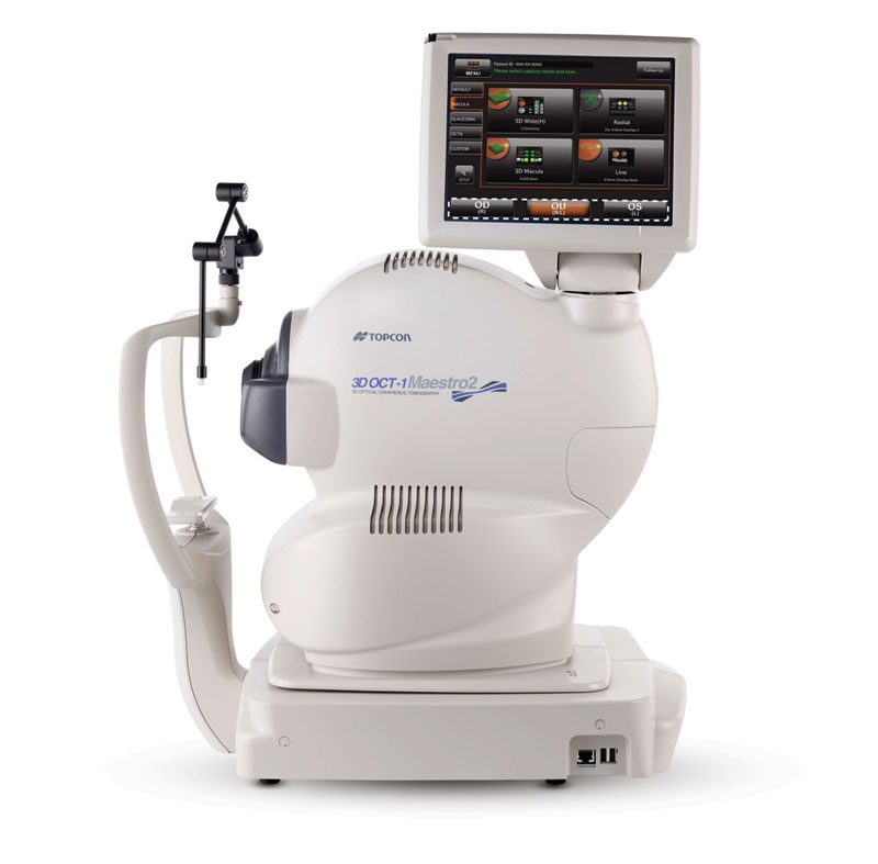
Practices that want a choice of imaging modalities in one device when documenting retinal disease can now consider the new Maestro2 from Topcon. A successor to the company’s 3D OCT-1, the new device can capture high-resolution non-mydriatic color fundus photography and conventional OCT or OCT angiography scans, the company explains. This multimodal system also now offers the Hood Report for better structure/function analysis of glaucoma, allowing easier comparison of RNFL scans with visual field defects shown on perimetry. It features a 360° rotating touchscreen, a small footprint and space-saving design, according to Topcon.
Clinical benefits described in a press release include easy image storing and sharing, precise repeatability in follow-up scans for better tracking of disease progress, and an extensive portfolio of reports for macular, anterior segment and glaucoma applications. For photography of the peripheral retina, the Maestro2 offers nine preset fields to choose from and the ability to manually control fixation; the device will then create a mosaic image of the fundus. A cataract mode adjusts the scanning position to compensate for opacity and a ‘live fundus’ view provides a real-time view of the retina for easier visualization of the optic disc or retinal vessels when determining OCT scan location.
Visit www.topconmaestro.com for more information.
