A 53-year-old Hispanic male presents complaining of blur at distance and near, headaches after work, and double vision while reading. He reports that when the double vision occurs, he feels eyestrain around his temples and feels as if an eye turns outward (although he is unsure which eye). How would you approach this case?
Diplopia is most commonly a symptom of eye misalignment. Alternatively, it may occur due to an issue as simple as a change in refractive error. But, in a worst-case scenario, diplopia may be the first sign of a muscular or neurologic disorder.
So, any complaint of diplopia must be a cause for concern.
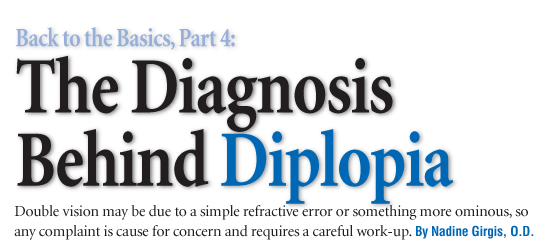
Exophthalmos is one possible finding in patients who present with thyroid disease, one of several possible diagnoses in patients who present with diplopia. Courtesy: Christopher Lievens, O.D.
History
A thorough history, while important for any comprehensive eye examination, can really help determine the diagnosis in patients with diplopia. Your history should include the following questions:
Is the diplopia monocular or binocular? This is the first question you should ask.1 The easiest way to do this is to ask whether the diplopia goes away if the patient closes one eye. Binocular diplopia goes away when either eye is closed. Monocular diplopia, however, goes away with only one particular eye closed, not either.2
Monocular diplopia is rare and is most often due to an optical aberration that results in multiple images in the diplopic eye. Causes of monocular diplopia include: corneal distortion or scarring (e.g., keratoconus), subluxation of the crystalline lens or an intraocular lens, vitreous abnormalities, multiple openings of the iris (polycoria), and retinal conditions (e.g., macular scarring or distortion).1 So, its no surprise that monocular diplopia can dissipate if a pinhole is used.3
Binocular diplopia most often occurs when the eyes are not aligned, and there are noncorresponding points on the two retinas stimulated. The binocular systems fusional capacity is disrupted, and a single image cannot be maintained. Changes in the optical function of the eye, which create aniseikonia, may also cause binocular diplopia, though much less frequently. Aniseikonia can occur secondary to refractive error, refractive surgery, or cataract surgery. The difference between these two types of diplopia: one object appears in two different locations vs. one object that appears differently (in size) in each eye.1,2,4
Is the diplopia constant or intermittent? Causes of intermittent diplopia include thyroid disease, myasthenia gravis, multiple sclerosis, and exo/eso breakdown. Causes of constant diplopia include cranial nerve palsy, thyroid or myasthenia gravis cases, and post-surgical cases. The most frequent surgical case that can result in diplopia is cataract surgery.
Is the diplopia present at distance, near or both? Many diagnoses can be ruled out if the diplopia is present only at a certain distance. This is important in cases such as divergence excess or convergence insufficiency/intermittent alternating exotropia at near.
What is the direction of the diplopia? Is it horizontal, vertical or torsional? The patient should explain what he or she sees after this question.
Causes of horizontal diplopia include thyroid eye disease (Graves disease or ophthalmopathy), internuclear ophthalmoplegia (INO), one-and-a-half syndrome and convergence insufficiency. Vertical diplopia may be a sign of thyroid eye disease, cranial nerve palsy, skew deviation, or Browns syndrome. Skew deviation and superior oblique, and inferior oblique palsy can all cause torsional diplopia.
How long has it been since the onset of diplopia?
Has the diplopia progressed or remained stable? Certain diagnoses are more likely to be progressive, such as multiple sclerosis, myasthenia gravis, and thyroid disease. Others occur suddenly and remain stable, such as cranial nerve palsy.
Have you ever had diplopia in the past? If so, is there anything that helps resolve the diplopia, such as looking in a certain direction or tilting the head?1 This question helps the doctor determine the diagnosis because some diagnoses, such as myasthenia gravis and thyroid disease, can produce multiple diplopic episodes. Others, such as cranial nerve palsies and INO, are generally one time occurrences.
As the patient answers each question, ask follow-up questions to supplement the incoming information.
Keep in mind that diplopia is less common in children than adults for several reasons. The most common is the lack of age-related understanding. If a child is asked if he/she sees double, he/she may not fully understand the question. Also, childrens visual systems are still maturing; therefore, they can more easily suppress multiple images than adults visual systems.
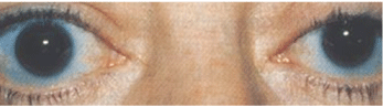
Ptosis and ocular misalignment can be clues to myasthenia gravis (MG). Some 48% to 53% of patients with MG initially manifest with only ocular MG. Courtesy: Paul C. Ajamian, O.D.
Supplementary Testing
Overall, the history takes time, but it helps you decide which tests to order and which diagnoses to consider.1 As you gather more information, tailor supplemental testing and referral to the possible working diagnoses.1
Besides the basic comprehensive eye examination, additional testing should include versions and ductions (forced ductions if possible). Parks three-step test (for vertical diplopia) and Hess Lancaster or red lens test (for horizontal diplopia) to help identify the paretic muscle. Realize, however, that these tests may yield inconclusive results if multiple muscles are involved, as with cranial nerve (CN) III palsy.
Not all cases of diplopia require a medical work-up, but you must perform exhaustive testing if a patient presents with diplopia and you choose not to refer.
Another consideration in the decision to refer a patient: comitancy. Often, the muscles involved secondary to a disease process or issue underact more in one direction than another, causing noncomitancy. The deviation will be larger in one of the nine fields of gaze vs. the other gazes. A longer-standing deviation is likely to be comitant; the magnitude of the deviation remains about the same in all nine gazes.
If a referral is warranted, the primary medical referrals should be to internal medicine; endocrinology; ear, nose, and throat; neurology; or neuro-ophthalmology. When extra testing is necessary, a full blood work-up should be requested. Additional testing includes an Enlon (formerly Tensilon) test to exclude myasthenia gravis, and Total T3/total triiodothyronine (T3), Total T4/total thyroxine/serum thyroxine (T4), and thyroid-stimulating hormone (TSH) with CT or MRI to rule out thyroid disease as well.
If there are multiple cranial nerve palsies, a full work-up is warranted given the higher likelihood of life threatening processes.5
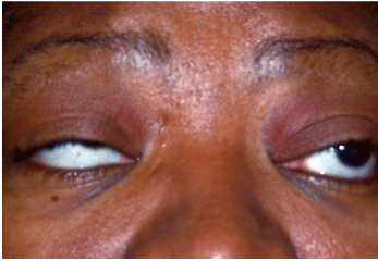
This patient with internuclear ophthalmoplegia presented with sudden onset of diplopia. On right gaze (top), he can abduct the right eye and adduct the left. On attempted left gaze (bottom), he shows decreased ability to adduct the right eye past the midline.
Causes of Diplopia
Courtesy: Andrew S. Gurwood, O.D.
There are a number of non-mechanical or neurological causes for diplopia. These include:
Uncorrected or undercorrected refractive error. Changes in refractive error can cause fixation switch diplopia, which occurs in patients with a history of strabismus. The change in refractive error or prescription causes fixation with the nondominant eye, resulting in diplopia. This can occur with monovision contact lenses, IOL implants, or following refractive surgery. Other optical causes can be related to optical centration or the switch from flat-top bifocal to progressive addition lenses.2,4
Thyroid eye disease (Graves disease or ophthalmopathy). This autoimmune disease is the most common cause of horizontal or vertical diplopia in adults. It also is the most common cause of exophthalmos in adults, and can present with other signs and symptoms, such as eyelid retraction, eyelid lag in downgaze, chemosis, eyelid swelling, foreign-body sensation, photophobia and decreased vision. Eyelid retraction is the most common sign; it occurs in 90% of patients with thyroid disease.6 Also, 60% have exophthalmos, and 40% have restrictive extraocular myopathy.7
Graves disease occurs secondary to hyperthyroidism, but ocular signs and symptoms (unilateral or bilateral) are not always present during this component of the disease.
Graves disease has a variable, but progressive course. Signs and symptoms can also occur after treatment or even in patients who are euthyroid (no lab signs of thyroid disease).
The inferior rectus muscle (limitation of elevation) is generally the first muscle involved. The medial rectus and superior rectus muscles sequentially are next in line of involvement. These patients commonly exhibit a hypotropia and esotropia; exotropia is rarely seen. Forced ductions, when performed, are positivea finding that differentiates this condition from many others. Extraocular muscles look enlarged on CT or MRI. TSH is below normal, while T4 and T3 are typically elevated.3,8,7-14
Myasthenia gravis (MG). This autoimmune disease is caused by a disorder of neuromuscular junction transmission that results in variable weakness of voluntary muscles. Some 48% to 53% of patients with MG initially manifest with only ocular MG, a variant that involves the extraocular, levator, and orbicularis muscles.15
MG can masquerade as many conditions, which affect any muscle or group of muscles. It may present as a hypertropia, esotropia or exotropia, or it can mimic any number of neurologic conditions. Additional signs and symptoms of ocular MG include ptosis and oculomotor weakness. There is no pupil involvement with MG.
MG is characterized by the variability of the signs and symptoms. These tend to worsen toward the end of the day when the patient is tired or after some strenuous activity. The Enlon test should be performed to help confirm diagnosis.1,3,9,16,17
Cranial nerve (CN) disease. Many different signs and symptoms accompany the diplopia complaint when the cranial nerves are involved secondary to any type of disease process.
CN III. The third cranial nerve can be partially or totally involved, and therefore can present with different signs and symptoms. Complete CN III involvement means the eye is ptotic with pupil involvement and that the eye cannot elevate or depress (i.e., the eye is down and out). If the pupil is spared and the patient complains of headaches and/or pain around the orbit, he or she most likely has microvascular disease (most commonly diabetes). Microvascular disease rarely affects more than one cranial nerve.
If the CN III palsy is partial, the eye will be ptotic and wont elevate if the superior division is involved or there is paresis of depression and adduction with a large, nonreactive pupil if the inferior division is involved.
CN III palsy can be congenital, due to birth trauma, or a neurologic syndrome. When it manifests in childhood, it is most commonly secondary to postviral infection, trauma or tumor (i.e., pontine glioma). The most common cause for CN III palsy in adults is microvascular disease, such as hypertension or diabetes, but it can also occur secondary to an aneurysm, trauma, or tumor.1,3,12
CN IV. With CN IV palsy, vertical diplopia most commonly manifests in adulthood secondary to decompensation of a congenital palsy. CN IV palsy can also be acquired.
It is important to differentiate between the two, because an acquired CN IV palsy is more cause for concern and possibly related to a disease process. Congenital CN IV palsy manifests with a consistent head tilt to the opposite side of the affected muscle since childhood. Also, there will be no complaint of image tilting, even though there is most definitely cyclotropia due to the fact that incyclotorsion is an action of the superior oblique. The patient will have large vertical fusional amplitudes and negative forced ductions.
Noncongenital CN IV palsy most commonly occurs secondary to trauma and microvascular disease, but in 30% of patients, it is idiopathic.8,5,18
When a patient has vertical diplopia, the Parks three-step test should be administered. To begin with, perform cover test in primary gaze. Which eye is the hypertropic eye? Then, perform cover test in left and right gazes (head turn). In which gaze is the hyper larger? Finally, with right and left head tilt, in which direction is the hyper larger?
At this point, a specific affected muscle can be determined if there is only one muscle causing the hypertropia. If there are multiple muscles involved, then the Parks three-step test will not help to single out the affected muscle.
CN VI. CN VI palsy presents as a noncomitant esodeviation that is worse in the field of gaze of the weak lateral rectus. The findings are negative on forced ductions testing.
CN VI palsy can be acquired at any age. If acquired in childhood, the most common cause is a tumor (specifically pontine glioma). Post-viral infection is the second most common cause.
Young adults have a different likelihood relative to children. Pontine glioma is not as common in this age group, and CN VI palsy is secondary to miscellaneous causes in this age group. Adults who present with CN VI palsy are more likely to acquire it secondary to trauma, microvascular disease, or multiple sclerosis.3,5
Case Report: Double Vision While Reading A 53-year-old Hispanic male presents complaining of blur at distance and near, headaches after work, and double vision while reading. He reports that when the double vision occurs, he feels eyestrain around his temples and feels as if an eye turns outward (although he is unsure which eye). His medical history is unremarkable.
Internuclear ophthalmoplegia (INO), one-and-a-half syndrome, and convergence insufficiency. These are three frequently observed causes of horizontal diplopia.
Upon examination, all pretests were normal until cover test was performed. At distance, with best correction, the patient had 16.00 prism diopter (PD) exophoria at near, and with an add, he had a 25.00PD intermittent alternating exotropia. Near point of convergence (NPC) was 13cm, and recovery was 25cm after three attempts. Also, base-out (BO) vergences were reduced at break and recovery.
Visual acuity was 20/20- O.U. at distance and near with a prescription of +0.75 -1.00 x 090 O.D. and +0.50 -0.75 x 085 O.S. and an add of +1.75. All anterior segment findings were within normal limits, and posterior segment findings lead to a primary diagnosis of glaucoma suspect.
We also diagnosed convergence insufficiency secondary to the larger exophoria/exotropia at near and reduced NPC and BO vergences.
We prescribed prism to alleviate some of the strain at near and eliminate diplopia. The prescription, given for near only, included prism of magnitude 3BI O.D. and 2BI O.S.
We instructed the patient to return one month later to follow up on the diplopia complaint, signs and symptoms, and do some further glaucoma testing, but he did not return to the clinic and was subsequently lost to follow up.
INO involves adduction weakness on the side of a lesion to the medial longitudinal fasciculus (MLF) and monocular nystagmus of the opposite abducting eye. It can be monocular or binocular (lesions of MLF-WEBINO, or wall-eyed bilateral INO). When binocular, the patient appears wall-eyed because each eye sits out laterally. Convergence is usually preserved, and diplopia occurs secondary to the lack of full range of eye movement.
One-and-a-half syndrome is a lesion of the MLF and abducens nucleus or paramedian pontine reticular formation (PPRF), so the affected eye cannot abduct or adduct. It is an INO plus, hence the name one-and-a-half syndrome.3,19-24 Both INO and one-and-a-half syndrome are most commonly acquired secondary to multiple sclerosis, tumor, or vascular disease.5
Convergence insufficiency, by definition, is primarily characterized by a larger exophoria (or exotropia) at near than at distance. Decompensation is possible in a previously stable eye misalignment as the patient ages. This occurs with congenital CN IV palsy and can occur with convergence insufficiency as well, in both cases, presenting as diplopia.
Skew deviation and Browns syndrome. These are often observed causes for vertical diplopia. Skew deviation can be comitant or noncomitant, but is an acquired vertical misalignment due to supranuclear dysfunction secondary to brainstem or cerebellar disease processes. Therefore, either eye is hyper, but the specific muscle cannot be determined by Parks three-step test. The patient also has multiple associated brainstem and cerebellar signs and symptoms, such as ataxia, nystagmus, confusion, and memory impairment.
Browns syndrome is a deficiency of elevation of an eye as it adducts. Its a mechanical restriction of the superior oblique tendon sheath; therefore forced ductions testing will be positive. Browns syndrome can be confused with an inferior oblique paresis, but inferior oblique paresis has negative forced ductions. The patient with Browns syndrome is hypotropic in primary gaze, and this condition is often congenital.1,5,8,9,25-34
Cataract surgery. Patients may complain of diplopia following cataract surgery due to four main reasons: surgical trauma to extraocular muscles or soft tissue around the muscles (most common), poor intraocular lens centration, a pre-existing condition (e.g., thyroid eye disease) with masked misalignment due to cataract, or conditions due to prolonged cataract occlusion (i.e., sensory deprivation).4,35
By no means is this an exhaustive list of causes for horizontal or vertical diplopia. Consider that any brainstem or midbrain issue or disease can cause diplopia.
Optical Treatment Options
Many treatment options can help resolve the diplopia, whether it be vertical or horizontal. An updated prescription should always be the first treatment.
Other options include nondisposable soft or gas-permeable contact lenses, which, specially ordered, can have small amounts of vertical prism in the lens.36 Monovision is another option.37 There are no contact lenses that contain horizontal prism due to feasibility issues.
Prism is often used and can be ground in or stuck onto the lens (Fresnel prism) in a pair of glasses. Fresnel prism is available in very small magnitudes from 1.00 prism diopter (PD) to large magnitudes of 40.00PD.
When there are no other optical treatment options, use occlusion. It is achieved by degrading the image of one eye, thereby eliminating monocular or binocular diplopia. Options for doing so include clear lacquer on the lens, translucent tape, a high plus lens, Bangerter filter, frosted lens or patching.1,2,38
Diplopia may occur secondary to something as simple as uncorrected refractive error or as complicated as brainstem disease in conjunction with multiple systemic signs and symptoms. As practitioners, we must keep in mind that diplopia is often an ominous sign and can be frightening to a patient.
It is very important to perform a good history along with multiple adjunct tests to determine if the diplopia is secondary to a systemic issue, which needs to be referred to a specialist, or can be managed by the optometric practitioner.
Dr. Girgis is an assistant professor at Nova Southeastern University College of Optometry in
1. Wessels IF. Diplopia. Available at: www.emedicine.com/ oph/topic191.htm (Accessed April 9, 2008).
2. Kushner BJ. Recently acquired diplopia in adults with long-standing strabismus. Arch Ophthalmol 2001 Dec; 119(12):1795-801.
3. Brazis PW, Lee AG. Acquired binocular horizontal diplopia. Mayo Clin Proc 1999 Sep;74(9):907-16.
4. Kushner BJ. Fixation switch diplopia. Arch Ophthalmol 1995 Jul; 113(7):896-9.
5. Friedman N, Pineda R, Kaiser P. The
6. Durairaj VD. Clinical perspectives of thyroid eye disease. Am J Med 2006 Dec;119(12):1027-8.
7. Bartley GB. The epidemiologic characteristics and clinical course of ophthalmopathy associated with autoimmune thyroid disease in
8. Brazis PW, Lee AG. Binocular vertical diplopia. Mayo Clin Proc 1998 Jan;73(1):55-66.
9. Rhee DJ, Pyfer MF. The Wills Eye Manual: Office and Emergency Room Diagnosis and Treatment of Eye Disease. 3rd ed.
10. Bartley GB, Gorman CA. Diagnostic criteria for
11. Bartley GB, Fatourechi V, Kadrmas EF, et al. The incidence of Graves ophthalmopathy in
12. Bartley GB, Fatourechi V, Kadrmas EF, et al. Clinical features of Graves ophthalmopathy in an incidence cohort. Am J Ophthalmol 1996 Mar;121(3):284-90.
13. Bartley GB, Fatourechi V, Kadrmas EF, et al. Chronology of Graves ophthalmopathy in an incidence cohort. Am J Ophthalmol 1996 Apr;121(4):426-34.
14. Vargas ME,
15. Brazis PW, Lee AG. Acquired binocular horizontal diplopia. Mayo Clin Proc 1999 Sep;74(9):907-16.
16. Bever CT Jr, Aquino AV, Penn AS, et al. Prognosis of ocular myasthenia. Ann Neurol 1983 Nov;14(5):516-9.
17. Weinberg DA, Lesser RL, Vollmer TL. Ocular myasthenia: a protean disorder. Surv Ophthalmol 1994 Nov-Dec;39(3):169-210.
18. Kanski JJ. Clinical Ophthalmology, 4th ed.
19. Sharpe JA,
20. Wall M, Wray SH. The one-and-a-half syndromea unilateral disorder of the pontine tegmentum: a study of 20 cases and review of the literature. Neurology. 1983 Aug;33(8):971-80.
21. Strominger MB, Mincy EJ, Strominger AI, Strominger NL. Bilateral internuclear ophthalmoplegia with absence of convergent eye movements. Clinicopathologic correlation. J Clin Neuroophthalmol 1986 Mar;6(1):57-65.
22.
23. Flitcroft DI, Saidlar CA, Stack JP, Eustace P. A proposed neuroanatomical and neurophysiological basis for WEBINO [abstract]. Neuroophthalmology. 1996;16:280. Abstract 46.
24. Komiyama A, Takamatsu K, Johkura K, et al. Internuclear ophthalmoplegia and contralateral exotropia: nonparalytic pontine exotropia and WEBINO syndrome. Neuroophthalmology 1998;19:33-44.
25. Spector RH. Vertical diplopia. Surv Ophthalmol 1993 Jul-Aug;38(1):31-62.
26. Allerand CD. Paroxysmal skew deviation in association with a brain stem glioma: report of an unusual case. Neurology 1962 Aug;12:520-3.
27. Hedges TR 3rd, Hoyt WF. Ocular tilt reaction due to an upper brainstem lesion: paroxysmal skew deviation, torsion, and oscillation of the eyes with head tilt. Ann Neurol 1982 May;11(5):537-40.
28. Straube A, Buttner U, Brandt T. Recurrent attacks with skew deviation, torsional nystagmus, and contraction of the left frontalis muscle. Neurology 1994 Jan;44(1):177-8.
29. Keane JR. Ocular skew deviation. Analysis of 100 cases. Arch Neurol 1975 Mar;32(3):185-90.
30. Keane JR. Alternating skew deviation: 47 patients. Neurology 1985 May;35(5):725-8.
31. Brandt T, Dieterich M. Pathological eye-head coordination in roll: tonic ocular tilt reaction in mesencephalic and medullary lesions. Brain 1987 Jun;110 (Pt 3):649-66.
32. Moster ML,
33. Halmagyi GM, Brandt T, Dieterich M, et al. Tonic contraversive ocular tilt reaction due to unilateral meso-diencephalic lesion. Neurology 1990 Oct;40(10):1503-9.
34. Brandt T, Dieterich M. Skew deviation with ocular torsion: a vestibular brainstem sign of topographic diagnostic value. Ann Neurol 1993 May;33(5):528-34.
35. Hunter DG, Lam GC, Guyton DL. Inferior oblique muscle injury from local anesthesia for cataract surgery. Ophthalmology 1995 Mar;102(3):501-9.
36. Lindsay RG, Crock GW. A bitoric rigid contact lens with base down prism to eliminate binocular vertical diplopia. Clin Exp Optom 2005 Jan;88(1):55-7.
37. London R. Monovision correction for diplopia. J Am Optom Assoc 1987 Jul;58(7):568-70.
38. Kirschen D, Flom MC. Monocular central-field occlusion for intractable diplopia. Am J Optom Physiol Opt 1977 May; 54(5):325-31.
