 |
| Accumulation of beta amyloid macroscopically induces a wide range of vascular abnormalities, as previous research suggests, including vascular attenuation, vessel tortuosity, narrowed veins, reduced branching complexity and increased width of vessels. Photo: Carolyn Majcher, OD, and Susan Ly Johnson, OD. Click image to enlarge. |
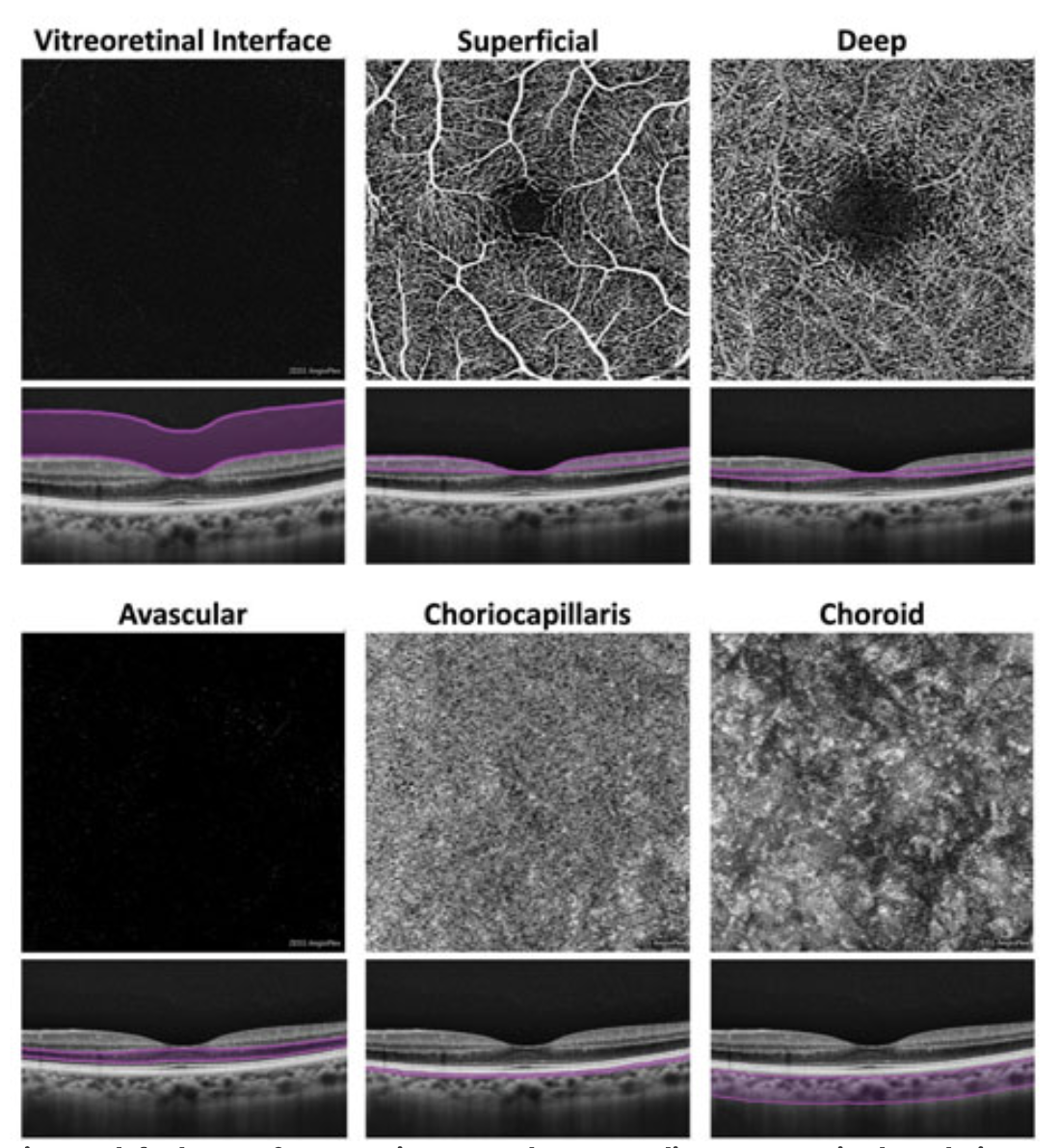
One new study outlined how the eye may be connected with neurodegenerative disease. Specifically, retina and choriocapillaris vascular structure characteristics were analyzed to look for simple, reliable biomarkers of Alzheimer’s disease. Since the brain and retina share embryologic origins and are affected through similar vascular changes, the study researchers thought OCT angiography (OCT-A) of these eye structures might reveal telling changes.
OCT-A was performed on 18 patients with early Alzheimer’s disease and 18 age-matched controls. All participants also underwent neurologic and ophthalmic examinations. What they found was that the choriocapillaris exhibited a significant flow area reduction of the choriocapillaris in the Alzheimer’s disease group. Subsequently, early Alzheimer’s may impair circulation of the structure.
The authors do mention that the choriocapillaris does not share the same embryologic origins as with the retina. However, its close relation to responsibility for nourishment and metabolism of retinal photoreceptors may make it susceptible to retinal damage from Alzheimer’s disease. Postmortem studies reveal beta amyloid plaques aggregated in retinal vessels of patients with Alzheimer’s, thus the vascular structure and fluid dynamics of the choriocapillaris may be vulnerable to damage from this kind of accumulation, even in the choroid, leading to possible modification of its anatomy and physiologic flow.
The authors continue that they found a trend of vessel density reduction of the superficial capillary plexus and another trend, although statistically insignificant, of reduced vessel density of the deep capillary plexus in mild cognitive impairment, which is in line with previous reports. One report describes these results as the potential result from direct beta amyloid accumulation inside vessel walls, causing loss of microvasculature and consequent reduction of vessel density through a local inflammatory process.
The early patients with Alzheimer’s included in this study and preferential involvement of the superficial capillary plexus is consistent with another prior report finding early involvement of the inner retina and superficial capillary plexus. Outer retina involvement was only observed with late impairment.
Based on their results and corroborated by similar studies findings, the authors stated “it has been hypothesized that OCT-A data from patients with Alzheimer’s are measures that could be used as an alternative biomarker to those currently available that may allow more easily accessible diagnosis and follow-up parameters for the disease, applicable even on a large scale.”
They did recognize, though, that “improved software for OCT-A devices is necessary, especially regarding choriocapillaris flow area assessment, and further studies are warranted to better understand the retinal and choroidal vascular changes in patients with Alzheimer’s disease to possibly use such values as biomarkers in clinical practice.”
Di Pippo M, Cipollini V, Giubilei F, Scuderi G, Abdolrahimzadeh S. Retinal and choriocapillaris vascular changes in early Alzheimer disease patients using optical coherence tomography angiography. J Neuroophthalmol. June 23, 2023. [Epub ahead of print]. |
