 |
|
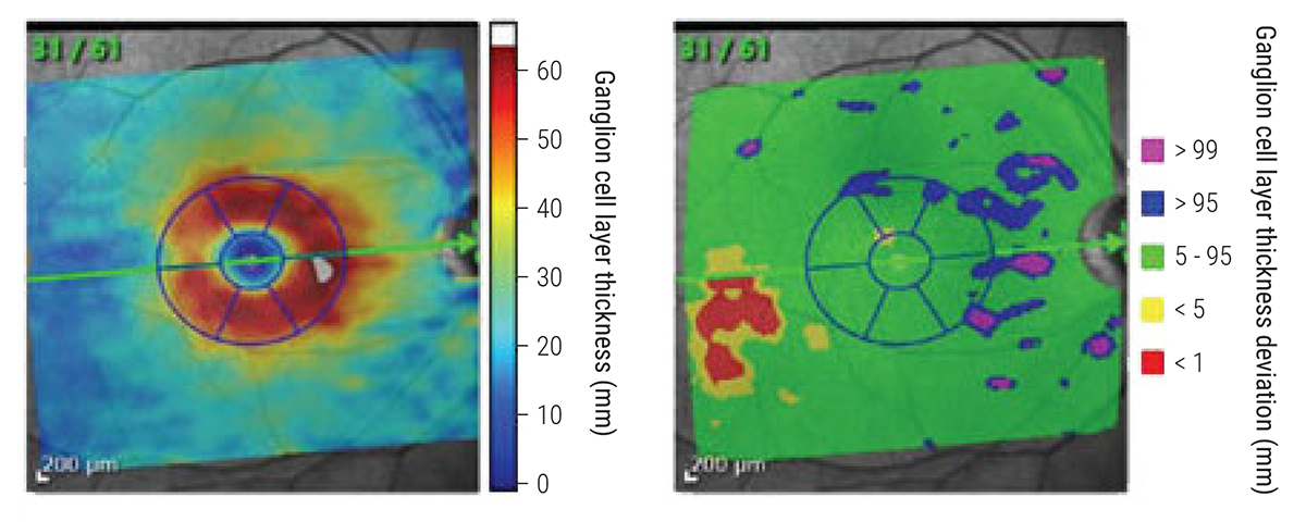
GCL thickness was more likely to demonstrate change compared with IPL in glaucoma suspects. Photo: James Fanelli, OD. Click image to enlarge. |
The current paradigm for structural evaluation of glaucoma with OCT devices is mainly based on thickness measurement as a biomarker for damage to various structures in the posterior pole of the eye such as the retinal nerve fiber layer, optic nerve head and inner macular layers. A recent study assessed and compared the rates of change of the inner plexiform layer (IPL) and ganglion cell layer (GCL) thickness in two cohorts of patients with suspected or established glaucoma to better delineate the respective changes in these two layers over time at different stages of the disease. The researchers found that, in a group of eyes with suspected glaucoma, GCL thickness declined more rapidly than IPL with no indication for preferential IPL loss.
The study consisted of 64 suspected glaucoma eyes (46 patients) and 112 established glaucoma eyes (112 patients) with ≥two years of follow-up and ≥three macular OCT scans. GCL and IPL superpixel thickness measurements were exported. A Bayesian hierarchical model with random intercepts, slopes and residual variances was used to estimate rates of change in individual superpixels. Normalized rates of change and proportions of superpixels with significantly negative and positive GCL and IPL rates of change were compared within the groups. Average follow-up time and number of scans were 3.5 years and 4.2, respectively, in the glaucoma suspect group and 3.6 years and 7.3, respectively, in the established glaucoma group.
In the glaucoma suspect group, a patient sample with suspected or very early glaucomatous optic neuropathy showed faster rates of GCL thinning compared with IPL (-0.69 vs. -0.33), and a larger number of superpixels in the central macula demonstrated higher probability of faster GCL rate of change compared with IPL. On the other hand, in eyes with established glaucoma, the IPL displayed relatively faster rates of decline than the GCL (-0.47 vs. -0.28), and a greater number of macular superpixels had a faster IPL rate of change.
“It is possible that the IPL thinning may accelerate with worsening of glaucoma, which seems to contradict the possibility of early pruning of the retinal ganglion cell dendrites,” the researchers wrote in their paper. “Early pruning of the dendritic arbor as manifested by thinning of the IPL cannot be corroborated in human glaucoma with the current resolution of OCT imaging.”
Mohammadi M, Su E, Chew L, et al. Comparison of ganglion cell layer and inner plexiform layer rates of change in suspected and established glaucoma. Am J Ophthalmol. December 11, 2022. [Epub ahead of print]. |
