Editor’s Note: As part of our “Year in Review” retrospective, we’ve selected the top 30 news stories of the year and are re-sharing them as we close out 2022. Follow along as we count down to number 1!
This story was originally published on October 28, 2022.
 |
|
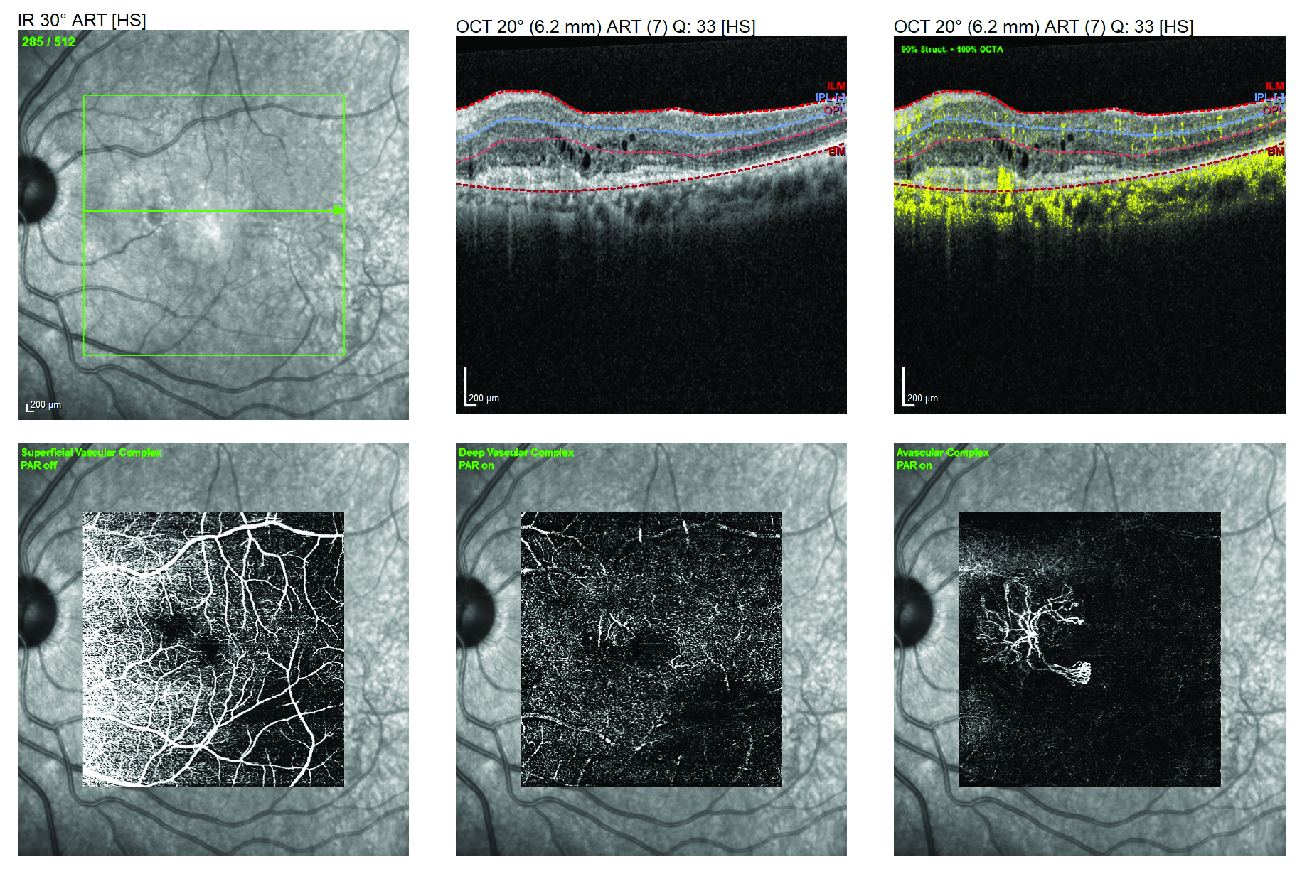
COPD patients show vascular differences on OCT-A. Photo: Lori Mandy Pennington, OD. Click image to enlarge. |
Although COPD primarily affects the lungs, the natural course of the disease seems to also encompass tissues of the eye, as indicated by one recent study. The work aimed to record retinal nerve fiber layer (RNFL) thickness and vascular density alterations in the retina and optic disc caused by the disease. Effects on the tissues were more pronounced in cases of severe COPD, and additional pronounced effects were seen in the areas of the deep capillary plexus and the radial peripapillary capillary plexus.
The prospective study included a total of 66 COPD patients (classified as mild, moderate or severe) and 54 healthy age- and sex-matched individuals. OCT-A was used to measure RNFL thickness and foveal avascular zone area as well as vessel density in the superficial capillary plexus (SCP), deep capillary plexus (DCP) and radial peripapillary capillary plexus (RPCP).
Analysis showed that COPD patients of all three categories display no differences from controls in foveal avascular zone area or RNFL thickness. However, the severe COPD group did display significantly lower vessel density of the deep capillary plexus in all parafoveal quadrants. Additionally, the severe group displayed lower vessel density of the radial peripapillary capillary plexus, especially of the peripapillary region.
The researchers propose that the reduced vascular density they found in the SCP, DCP and RPCP of the severe COPD group may be the cause of vascular structural loss, atherosclerotic changes or chronic hypoxia and endothelial dysfunction caused by vasoconstriction. Since autoregulatory mechanisms control retinal blood flow, atherosclerotic changes, hypoxia or endothelial dysfunction could alter this blood flow by disrupting the autoregulation. Even further, almost all vascular density parameters had a positive correlation with partial pressure of oxygen levels, including the SCP, DCP and RPCP values.
Understanding the connection between COPD and ocular changes begins with inhaled irritants, usually cigarette smoke, causing COPD patients to release inflammatory mediators into circulation. Subsequently, vascular endothelial cells activate, and this causes the inflammatory process to ensue. Endothelin 1 (ET-1), increased by this this inflammatory response, is seen in urine of COPD patients and is linked to endothelial dysfunction. ET-1 has been documented to cause dose-dependent vasoconstriction in the microvasculature of the retina, bringing COPD back to the forefront of the problem, elucidating at least in part why there is a connection between the condition and observed microstructural changes.
While those most at risk primarily include those with severe manifestation of the disease, neither mild nor moderate cases of COPD should be ignored when looking at potential ocular effects of the retina and optic nerve. As the authors similarly mention, for everyone with the condition, “we believe that OCT-A is an important tool in the follow-up of COPD patients and can be used together with spirometry as a noninvasive method of monitoring progression in these patients.”
Incekalan TK, Safçƨ SB, Şimdivar GHN. Investigation of ocular microstructural changes according to disease severity in patients with chronic obstructive pulmonary disease. Can J Ophthalmol. 2022. [Epub ahead of print]. |
