 |
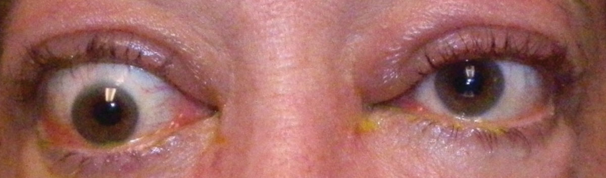
| Thyroid-associated ophthalmopathy affects numerous corneal parameters due to proposed mechanisms including tension exerted on the cornea through eyelid pressure and the forces derived from extraocular muscles exerted through the sclera. Photo: Bobby Saenz, OD. Click image to enlarge. |
The most common immune orbital disorder, thyroid-associated ophthalmopathy (TAO), also known as Graves’ eye disease, Graves’ ophthalmopathy or Graves’ orbitopathy, is characterized by a long list of clinical features, including upper eyelid retraction, erythema, edema, proptosis, corneal exposure and impaired vision. A primary sign that the disease has become moderate to severe is fibrosis of the orbital tissue, particularly the extraocular muscles. The forces from these muscles are then exerted through the sclera, which, accompanied by eyelid and ocular surface inflammation, may contribute to the corneal morphological and biomechanical changes that a recent study observed in TAO patients.
The small study, published in Cornea, enrolled 53 eyes with varying severities of TAO and 16 healthy eyes. Of eyes with TAO, 28 had active disease and 25 had inactive disease. All underwent complete ophthalmic examinations including magnetic resonance imaging. The researchers used Pentacam imaging to measure corneal topography and a Corvis ST tonometer to obtain biomechanical parameters. Next, they evaluated the correlations between corneal parameters, clinical activity score and NOSPECS score and calculated the diagnostic accuracy of corneal changes for active and severe TAO.
The following corneal parameters were increased in patients with TAO: back elevation, Corvis biomechanical index, tomographic and biomechanical index, stiffness parameter at the first applanation, deviation from normality in back elevation, relational thickness and overall deviation from normality. Conversely, significant decreases were noted in TAO patients in the smallest corneal thickness, maximum Ambrósio relational thickness and deformation amplitude.
In distinguishing mild TAO from moderate-severe TAO, the parameters showing the highest diagnostic capability were maximum Ambrósio relational thickness, Ambrósio relational thickness through the horizontal meridian and corneal thickness. The researchers noted in their paper that this finding “[indicates] that these parameters could serve as effective references for evaluating the severity of TAO.”
Disease activity also had an effect on various corneal parameters. A strong positive correlation was found between clinical activity score and back elevation. “The differences in corneal morphological and biomechanical parameters between the active and inactive groups provide evidence of the effect of inflammation on corneal changes,” the study authors noted.
While the pathogenesis and mechanism underlying these corneal changes in TAO patients are likely multifactorial, the researchers proposed a few possible explanations.
“Many inflammatory cytokines, such as IL-6, TNF-a, and TGF-b, have been found on the ocular surface of patients with TAO, especially those in the active stage,” they wrote in their paper. “The interactions of these inflammatory cytokines are well established in the molecular pathways involved in corneal development, inflammation and wound healing and lead to keratocyte apoptosis and decreased keratocyte cell density.”
The authors also emphasized that “the forces transmitted to the cornea through the sclera derived from the stiff extraocular muscles cannot be ignored. The corneal topography could be influenced by strabismus surgery through alteration of extraocular muscle tension or intraocular pressure.” They then pointed to a study that observed an influence of orbital soft-tissue fibrosis on corneal astigmatism, as well as lower corneal thickness and corneal biomechanical stability in moderate-severe TAO vs. mild TAO.
Though the present study included a small number of patients, its findings align with existing literature that shows corneal changes do present in TAO patients which researchers attribute to inflammation of the ocular surface. Additionally, this study found that these changes correlate with disease activity and severity. Based on these observations, the researchers conclude that “measurements of corneal morphological and biomechanical parameters could serve as references in evaluating TAO.”
Zhang T, Ye H, Xiao W, Chen R, Huasheng Y. Corneal morphological and biomechanical changes in thyroid-associated ophthalmopathy. Cornea. 2023;00-1-7. |
