 |
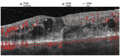
Researchers found that it was more beneficial to use OCT-A over other imaging methods when identifying and diagnosing patients with macular neovascularization. Photo: Steven Ferrucci, OD, and Jay M. Haynie, OD. Click image to enlarge. |
Practitioners with access to OCT-A have come to rely on it for the evaluation of retinal disorders with a vascular component, but protocols are still being developed for these newer applications. To better understand the relative merits of cross-sectional and en face OCT-A, researchers recently conducted a prospective cohort study comparing both and found that cross-sectional OCT-A not only yields high sensitivity and specificity in the detection of this condition but also improves diagnostic accuracy vs. en face OCT-A imaging alone.
The study included 102 eyes of 63 consecutive patients with various chorioretinal diseases who had subretinal hyperreflective material and/or pigment epithelial detachment on OCT, possibly corresponding to macular neovascularization in at least one eye. Enrolled patients underwent fluorescein angiography (FA), OCT and swept-source OCT-A.
The researchers evaluated FA images, the outer retina to choriocapillaris OCT-A en face slab, a manually modified en face slab and cross-sectional OCT-A for the presence of macular neovascularization. A combination of cross-section and en face OCT-A was also analyzed.
A sensitivity for the detection of macular neovascularization of 46.3% and a specificity of 93.4% was observed when using the automatically provided outer retina to choriocapillaris OCT-A en face slab. The manually modified slab showed a sensitivity of 78.1% and a specificity of 88.5%, the study authors reported. Comparatively, cross-sectional OCT-A resulted in a sensitivity of 85.4% and a specificity of 82%. When evaluating en face in combination with cross-sectional OCT-A, macular neovascularization was identified with a sensitivity of 82.9% and a specificity of 85.3%.
These findings demonstrate the high level of sensitivity and specificity provided by cross-sectional OCT-A, according to the study authors, who also noted that the decreased sensitivity for disease detection on automatically generated OCT-A en face images is a result of segmentation errors.
“Cross-sectional OCT-A is independent of the position of segmentation lines and may improve accuracy of image evaluation compared with en face images alone by identifying the macular neovascularization-suspicious area and without time-consuming manual correction of segmentation errors in daily routine,” the researchers concluded.
Siggel R, Spital C, Lentzsch A, et al. Optical coherence tomography angiography for the detection of macular neovascularization—comparison of en face versus cross-sectional view. Eye (Lond). January 6, 2022. [Epub ahead of print]. |
