 |
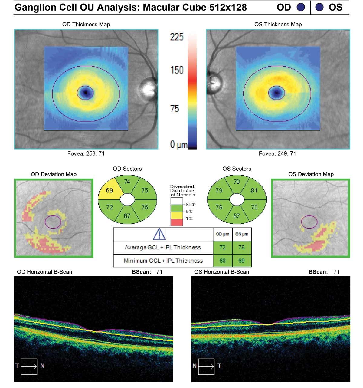
| Unlike brain volume measured by MRI, GCL thickness (shown here in a glaucoma patient) is not directly affected by inflammation, representing a valuable measure of neurodegeneration especially relevant for MS prognosis. Photo: Carl Zeiss Meditec. Click image to enlarge. |
The macular ganglion cell layer (mGCL) often holds clues related to systemic neurodegenerative diseases, and can be a useful biomarker of axonal degeneration in multiple sclerosis (MS). The researchers of a recent study sought to develop a deep learning technique using mGCL thickness data to aid in MS diagnosis and prognosis.
The team evaluated the findings of a cross-sectional study comprising 72 MS patients and 30 controls for diagnosing disease and a 10-year longitudinal study of the same MS patients for predicting progression. The mGCL was measured with OCT.
The deep learning algorithm for MS diagnosis had an accuracy of 90.3%, and prediction of disease progression eight years later resulted in an accuracy of 81.9%. The researchers wrote in their paper for Acta Ophthalmologica that “a decrease in mGCL thickness could be one of the most promising biomarkers of MS-associated neurodegeneration.”
Looking at the statistical analysis between MS patients and controls in the study, MS patients displayed both lower macular volume and different mGCL thicknesses. The central fovea was the only area not significantly affected.
Of the many characteristics measured between patients and controls, the only significant difference was found in the Expanded Disability Status Scale (EDSS) over three years. EDSS scores in MS patients without disease progression were higher than for MS patients with progression. However, macular volume and mGCL thickness were both higher in MS patients with progression. The authors explained that this is consistent with prior research indicating axonal damage cumulatively occurs from MS onset, with retinal thinning occurring early in the disease process.
The authors noted that because many artificial intelligence models lack mGCL thickness as a measure to predict MS progression, they had to compare their model to those looking at retinal nerve fiber layer (RNFL) thickness instead. Their prior research using RNFL OCT metrics resulted in 95.8% accuracy for MS diagnosis and 91.3% accuracy for prognosis.
The team also noted that the EDSS is how disability progression is primarily measured; however, the scale lacks some nuance, incompletely reflecting neurodegenerative damage, not accounting for neuropsychological disability and producing a low sensitivity. They compare this scale against OCT, which offers the benefits of being noninvasive, cost-effective and fast while producing standardized, reliable, quantitative measurements.
The authors are hopeful OCT will make its way into mainstream MS management, concluding that “including the use of OCT technology in the treatment of MS would bring great benefits to clinicians, who would make early and comprehensive diagnosis and would select the most specific treatments with which to improve patients’ lives.”
Montolío A, Cegoñino J, Garcia-Martin E, Pérez del Palomar A. The macular retinal ganglion cell layer as a biomarker for diagnosis and prognosis in multiple sclerosis: a deep learning approach. Acta Ophthalmol. June 10, 2023. [Epub ahead of print]. |
