24th Annual Surgery ReportFollow the links below to read other articles from annual update on surgery: The Preoperative Ocular Surface Checkup Understanding the Role of IOL Optics in Postoperative Vision Complaints Corneal Compromise: How to Assess the Risk of Post-LASIK Ectasia |
Optometrists are the primary eye care providers for the majority of Americans, and ophthalmology services may be hours away for those in many rural areas.1,2 Therefore, it is incumbent on us to practice to the full scope of our licensure and training to provide our patients the best and most effective care we can. Depending on your state legislature, amending your skill set may encompass adding minor surgical procedures to your repertoire. Doing so will positively impact patients’ lives, providing them access to valuable services closer to home.
Adding surgical procedures is easier than you might expect. In fact, many optometrists already perform such tasks—foreign body removal, eyelash epilation, and dilation and irrigation of the puncta—on a daily basis. Broadening optometric privileges across the country is bringing these opportunities to more optometrists than ever. Optometrists in Alaska, Kentucky, Louisiana, Nebraska, New Mexico, Oklahoma, Oregon and Tennessee can perform minor surgical procedures. Idaho, Montana, North Carolina, North Dakota, Utah, Virginia, West Virginia, and Wisconsin allow the use of injectable drugs for diagnostic and treatment purposes.3-11
This article reviews the basic minor surgical procedures that you can perform in your office, though it is important to consult your state board about which are permissible under your state’s optometric law.
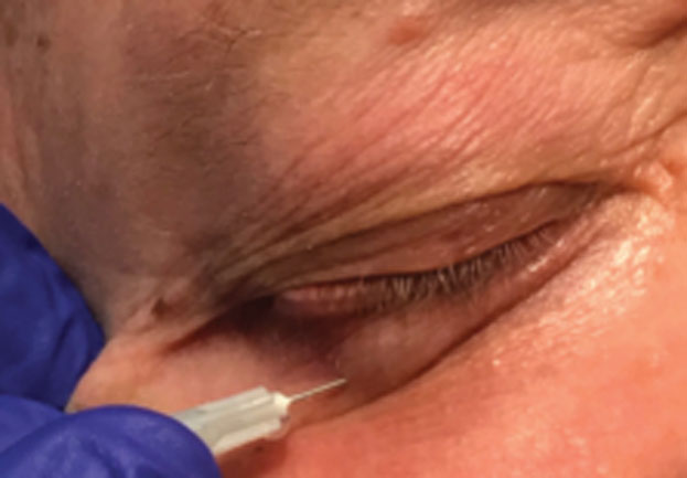 |
| Fig. 1. When injecting anesthesia, pull the eyelid taut and ask the patient to look away from the injection site. |
Medical History and Informed Consent
Best practices dictate acquiring a thorough medical history and obtaining informed consent from the patient prior to any procedure. The history should include any past or present medical conditions, drug and latex allergies and a list of all medications the patient is taking, including over-the-counter ones.
Pay special attention to any anticoagulants the patient is taking, such as aspirin, nonsteroidal anti-inflammatory drugs, warfarin, heparin, dipyridamole and clopidogrel. Anticoagulants increase the risk of bleeding and lengthen healing time. Consider speaking with the patient’s primary care provider to determine if the patient can suspend treatment of the anticoagulant to have the lesion removed.
It is also important to inquire about cardiovascular health, especially hypertension. Epinephrine, which is commonly added to anesthetics, should be used with caution in patients taking tricyclic antidepressants and beta-blockers. Epinephrine should not be used in patients with severe hypertension, hyperthyroidism and pheochromocytoma.12
Pregnancy is also an important screening question in all women of child-bearing age, as anesthetics may act as possible teratogens.12
Your informed consent discussion should give the patient the opportunity to ask questions and have them answered to their satisfaction prior to signing an acknowledgment of the risks and electing to proceed. Patients must be at least 18 and capable of independent decision-making to sign an informed consent. A parent or other legal guardian may do so for a minor or an adult incapable of making their own informed decisions.12
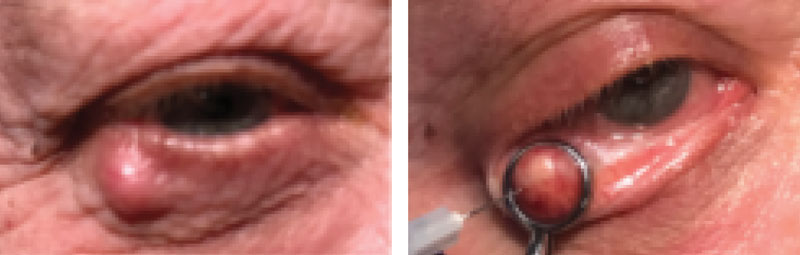 |
| Figs. 2 and 3. Before making an incision on the chalazion (left), carefully inject the anesthetic. You may use a chalazion clamp to isolate the lesion before injection (right). |
Prepare Anesthesia
Periocular injections are used for local anesthesia and/or administration of medication locally into affected tissue. Sterile, single-use disposable stainless-steel needles are used for both types of injections.13 Small-diameter short needles are the best choice for periocular tissues, as these provide maximum comfort and precise control.13 Most commonly used needles range from 3/8” to 2” in length and 27- to 30-gauge in diameter. Remember, the gauge of a needle is inversely proportional to its diameter—the higher the gauge, the smaller the diameter.13
There are several types of anesthetic of varying concentrations for use in periocular injections, but Xylocaine (lidocaine hydrochloride, Astra Zeneca) and Marcaine (bupivacaine hydrochloride, Pfizer) are the most common choices. Lidocaine has a shorter onset of action (two to four minutes) but only provides anesthesia for about one hour. While bupivacaine has a longer onset of action (10 minutes), its duration can range from four to six hours.13,14 Some practitioners find it useful to use a combination of both to obtain a faster onset of action with a prolonged duration.13,14
A helpful addition to the anesthetic is a vasoconstrictor, such as epinephrine, which works to minimize bleeding and prevent systemic absorption of the anesthetic.13 In practice, we prefer to use the prepared combination of 2% lidocaine with epinephrine 1:100,000. This provides a quick anesthesia that lasts well past the amount of time required for a minor lid procedure.
A drawback of local infiltrative anesthesia is discomfort due to the anesthetic’s low pH. While lidocaine’s falls between 5.0 and 7.0, preservatives added by the manufacturers lower the pH of lidocaine/epinephrine to anywhere from 3.3 to 5.5. Buffering with 8.4% sodium bicarbonate at a ratio of 1 to 10 lidocaine/epinephrine 1:100,000 or 1 to 15 lidocaine/epinephrine 1:200,000 will bring the pH close to the physiologic pH, thereby reducing pain with injection.12,13 Adding sodium bicarbonate also shortens the duration of action of epinephrine while speeding up lidocaine’s onset of anesthesia.12,13 Another simple step to increase comfort is to bring the temperature of the anesthetic up to body temperature prior to administration.15
Local anesthesia is not without potential side effects. Though rare, severe allergic reactions, contact dermatitis, lightheadedness, nausea, bradycardia, hypotension and seizures are possible.12 In addition, some patients can experience psychogenic attacks due to anxiety over the procedure or fear of needles. These types of attacks often elicit a vasovagal response, which can lead to syncope.12 For that reason, most injections are performed with the patient in a supine position.
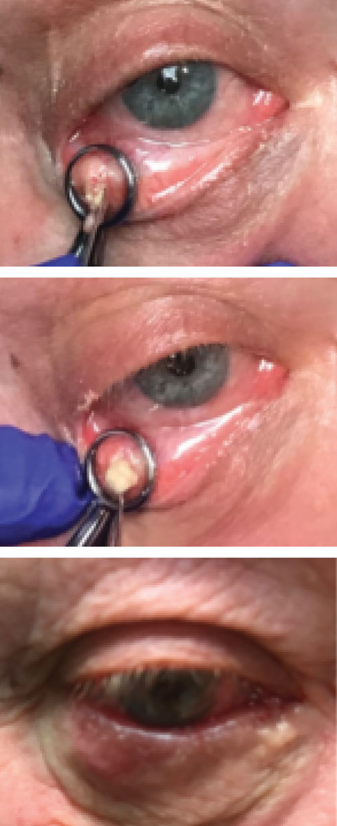 |
| Figs. 4-6. Granulomatous material will extrude from the lesion upon incision (top). Curette the chalazion (middle) upon incision. Eyelid immediately post-op (bottom) shows bruising from the procedure but overall improvement in cosmetic appearance. |
Handle Injections With Care
The two types of injections are subcutaneous and intradermal. Subcutaneous injections are how local anesthesia is administered—below the epidermis and dermis layers—while intradermal injections are given directly into the lesion.
Subcuteous injection for anesthesia. First, clean the top of the medication vial with an alcohol wipe. Prepare the syringe by drawing the solution from the vial using an 18-gauge needle. Then, change to a 27- to 30-gauge needle in preparation for administration. Some clinicians use a Jaeger plate or corneal shield to protect the globe during the injection. To do so, first instill ophthalmic proparacaine, then have the patient look in the opposite direction of the lid to be injected and insert the plate posterior to the lid and anterior to the globe. Pull the eyelid taut and ask the patient to look away from the site you are injecting. Let the patient know they will feel a needle stick, and, with the bevel of the needle facing up, insert the needle in a subtle stabbing motion at a 15-degree angle to the skin into the subcutaneous layer beneath the lesion (Figure 1).
Slowly inject the desired amount of anesthetic, creating a bolus under the skin, and then remove the needle slowly. Depending on lesion size and shape, it may be necessary to inject the lesion at additional sites. For increased patient comfort, it is best to go through the already anesthetized area for subsequent injections. Once the desired amount is injected and encompasses the lesion to be removed, place gauze over the area while applying gentle pressure and massage the anesthetic into the tissue. Make sure to test for numbness before beginning any procedure.13
Intradermal injection for chalazion. Administering 40mg/mL Kenalog (triamcinolone acetate, Bristol-Myers Squibb) provides a viable alternative to incision and curettage.8,13 In some instances, such as with children or when lesions are close to the lacrimal apparatus, steroid injection is actually the treatment of choice.13
Treatment can be performed with or without prior anesthesia. Injection approach can be made through the palpebral conjunctiva or the external eyelid. The transconjunctival approach is preferred in darkly pigmented individuals, as focal hypopigmentation can occur at an external injection site.13 A chalazion clamp is typically used to isolate the lesion and protect the globe.
The medication must be well-shaken before being drawn and needs to be injected soon after preparation to keep it from precipitating out of the solution. Draw 0.5mL of triamcinolone acetate into a syringe with a 25-gauge needle. Next, stand beside the patient, pull the lid taut and have the patient look opposite the area of injection. The angle to inject the needle will vary depending on the size of the chalazion but should be shallow enough to prevent full-thickness penetration of the eyelid and/or globe perforation. Insert the needle directly into the center of the lesion using a technique similar to the one described earlier for anesthesia. Remember that some resistance may be encountered due to the wall of the chalazion. While only about 0.05mL can be injected into a chalazion, some clinicians also elect to inject 0.1mL of additional steroid paralesionally to help achieve further, albeit more gradual, penetration.
Results from triamcinolone injections can be seen between one to four weeks. If the chalazion is not completely resolved at one month, you may give an additional injection or perform incision and/or curettage.13 The success rate of only one injection resolving a chalazion is approximately 80%.16
An important caveat regarding the injection of intralesional corticosteroid is the potential for central retinal artery occlusion due to the retrograde passage of particulate from a periorbital artery in the eyelid into the ophthalmic artery past the point of branching of the central retinal artery. When using needles with larger diameters, inject the steroid with as little force possible and know the anatomy of eyelid and adnexal vasculature in order to minimize this risk. Adult human periocular arterioles have average lumen diameters of 0.5mm.17 A 27-gauge needle has an external diameter of 0.41mm while a 25-gauge needle has a diameter of 0.52mm.18,19 While the risk of this occurring is low, there have been several documented cases of retinal artery occlusion following intranasal, forehead and eyelid corticosteroid injections.20
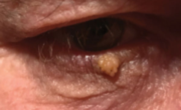 |
| Fig. 7. A squamous papilloma is a benign lesion that can be removed in the office. |
Become a Surgical Expert
These surgical treatment options require a lot of attention to detail in order to avoid complications, such as scarring. Get these techniques down pat and learn to use the appropriate tools correctly.
Incision and curettage of chalazia. The definitive yet more invasive treatment for chalazia is incision and curettage (Figure 2).19 This procedure may be selected as the initial treatment option due to its high success rate or when a chalazion has failed to respond to corticosteroid injections. Instill topical proparacaine to help increase patient comfort during the procedure. Then, inject the anesthetic around the lesion externally as described previously to numb the eyelid around the chalazion (Figure 3). After several minutes, test the area for aesthesia.
Once numb, evert the eyelid and place a chalazion clamp with the open ring surrounding the lesion on the palpebral conjunctiva and the plate against the outer eyelid. Allow the clamp to hang gently from the eyelid and rest on the forehead or cheek, away from the cornea. At this point, if so desired, more anesthetic can be injected into the chalazia through the palpebral conjunctiva while remaining anterior to the tarsal plate. A sterile surgical blade is then used to make a 3mm vertical incision over the chalazion, parallel to the meibomian glands. It is best to remain 2mm to 3mm away from the lid margin to prevent notching of the eyelid.
Upon incision, granulomatous material will often erupt through the opening (Figure 4). Next, insert the curette into the incision and begin to scrape and scoop the material from the chalazion (Figure 5). You may send some of this material for pathology if desired, but always send it in any case of recurrent chalazion due to the possibility of meibomian gland carcinoma.13,16
To prevent recurrence, ensure the entire sac surrounding the lesion is removed. Grasp the sac with toothed forceps and use surgical scissors to detach it from surrounding tissue if needed. Remove the clamp and hold gauze over the lid while applying gentle pressure for hemostasis. In case of recalcitrant bleeding, a disposable thermal cautery unit can be used. Instill ophthalmic antibiotic ointment, such as erythromycin or polysporin. Some prefer to use a steroid/antibiotic combination for anti-inflammatory coverage.
Prescribe an ophthalmic antibiotic for one week post-procedure. Be sure the patient understands that drainage may occur for several days. Educate the patient to return to the clinic if any signs/symptoms of infection occur and schedule the patient for follow-up in five to seven days to ensure adequate healing is obtained (Figure 6).13
Snip excision. Benign lesions such as squamous papilloma, seborrheic keratosis and verruca can be removed in-office by simple snip excision (Figure 7). Signs of benign lesions include even coloration, well-defined regular borders, lack of ulceration, no induration, a history of slow growth and maintenance of normal skin structures such as lashes and glands.16 We often find patients can tolerate snip excision without anesthesia, especially for pedunculated masses. If anesthesia is desired, inject the anesthetic at the lesion base.
Once the area is numb, grasp the mass with toothed forceps, pull it slightly away from its base and snip it free (Figures 8, 9 and 10). Light pressure with a small gauze pad for a few minutes usually stops any bleeding, but a disposable thermal cautery unit comes in handy if bleeding continues. Place the specimen in a formalin container suitable for transport to the laboratory for histologic evaluation.
Prior to releasing the patient, apply a prophylactic antibiotic ointment, such as erythromycin or polysporin, and advise them to keep the area clean and dry and apply the ointment three times daily for three days. If any signs or symptoms of infection develop, the patient should return to the office.13
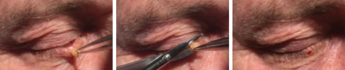 |
Figs. 8-10. When excising a squamous papilloma, isolate with foreceps to expose the base (left) and use surgical scissors to snip the lesion free at its base (middle). Note the minimal amount of blood seen after lesion removal with snip excision (right). |
Excision using the Ellman radiosurgical probe. Lesions that have a broader base and cannot be easily removed with a simple snip excision may be good candidates for removal with the Ellman Surgitron instrument. This device applies high-frequency radio waves to tissue to excite water molecules and generate heat, vaporizing the water and causing cell lysis. This creates a sterile cut with very clean edges. This type of surgical technique has the advantages of minimal blood loss and rapid healing.21
There are a few drawbacks to using the Ellman. Vaporization of the tissue produces vapor with a very strong odor and can release particles that could be inhaled (think verruca) into the air. Hence, it is important to use the compatible vacuum apparatus to remove any resultant vapor. This instrument must not be used with patients who have pacemakers, as it can interfere with the device’s function. It is also important to avoid using this instrument in the presence of flammable fumes or liquids.21
To perform a lesion removal using the Ellman device, have the patient in a supine position with their head against the exam chair’s headrest. The ground plate, necessary for transmitting the radio waves, can be placed between the patient’s shoulder and the chair. The lesion and surrounding area must be prepped with a betadine swab. After several minutes, administer the anesthetic. When the patient is sufficiently numb, lesion removal can begin.
Turn on the device and allow it to warm up for at least 30 seconds. Choose the appropriate waveform and power setting. Our practice tends to use the cut and coagulation waveform with a power setting of four, though there is slight variability to each unit, and you should take time to become accustomed to the various settings on your specific device. To activate the electrode, press down the footplate. Radio waves are most effectively transferred to tissue with a high water content, so keeping the lesion surface moist with sterile gauze saturated with sterile saline in between passes of the electrode facilitates effective lesion removal. Grasp and lightly pull pedunculated lesions with tissue forceps and then use a loop electrode to cut at the base of the lesion to remove in one piece. The remaining edges can then be cleaned with passes of the probe until a uniform appearance is achieved.
The other method, feathering, is helpful in broad-based sessile lesions and involves passing over the lesion with the electrode, removing it in layers using a brush-like motion, cleaning and re-wetting after a few passes. A ball electrode may be employed in the rare case of excessive bleeding. Once removal of the lesion is complete, clean the area with sterile saline to remove any betadine remaining. Apply a layer of topical ophthalmic antibiotic over the affected area.21,22 Again, have the patient apply antibiotic ointment three times per day for three to seven days to ensure proper healing.
Optometrists care for the eye and vision needs of more Americans than any other medical professional. We should seize this opportunity to provide the most efficient and state-of-the-art services and procedures possible, as taught in our professional schools and continuing education courses. Add these procedures to your professional skillset and you will be on your way to doing so.
Drs. Burress, Bendure and Kedzuf are all staff optometrists at the Ernest Childers VA Outpatient Clinic in Tulsa, OK, and adjunct professors at the Oklahoma College of Optometry.
1. Optometric surgical privileges improve access to care, ease financial burdens. Healio. www.healio.com/optometry/primary-care-optometry/news/print/primary-care-optometry-news/%7Bcb12a392-483f-41bb-8f2f-cfc3cfbc2083%7D/optometric-surgical-privileges-improve-access-to-care-ease-financial-burdens. Published January 2012. Accessed December 15, 2018. 2. An action-oriented analysis of the state of the optometric profession: 2013. Jobson Medical Information. 2013. www.aoa.org/Documents/news/state_of_optometry.pdf. 3. Governor Bill Walker signs HB 103 into law [news release]. Juneau, AK. Office of the Governor; July 11, 2018. akoa.org/news_manager.php ?page=14408. Accessed December 15, 2018. 4. Eisenberg J. Kentucky expands ODs’ scope of practice. Rev Optom. 2011;148(3):4-6 5. Louisiana Gov Jindal signs expanded scope of practice bill. AOA News. www.aoa.org/news/advocacy/louisiana-governor-jindal-signs-expanded-scope-of-practice-bill. Published June 2, 2014. Accessed December 15, 2018. 6. Kelly E. Nebraska increases scope-of-practice for ODs. Review of Optometry. www.reviewofoptometry.com/article/nebraska-increases--scope-of-practice-for-ods. Published May 15, 2014. Accessed December 15, 2018. 7. Murphy J. New Mexico passes minor surgery law. Review of Optometry. www.reviewofoptometry.com/article/new-mexico-passes-minor-surgery-law. Published May 4, 2007. Accessed December 15. 2018. 8. Fenelli J. Injection: the third method of drug administration. Rev Optom. 2012;149(1):32-40. 9. Oregon State Law. Chapter 683. Optometrists; Opticians. 2017 Edition. www.oregonlegislature.gov/bills_laws/ors/ors683.html. Accessed December 15, 2018. 10. Legislation in Tennessee to allow ODs to use injectable anesthetic. Review of Optometry. www.reviewofoptometry.com/article/legislation-in-tennessee-to-allow-ods-to-use-injectable-anesthetic. Published April 15, 2014. Accessed December 15, 2018. 11. RO staff. Virginia ODs enjoy expanded scope of practice. Review of Optometry. www.reviewofoptometry.com/article/virginia-ods-enjoy-expanded-scope-of-practice. April 11, 2018. Accessed December 15, 2018. 12. Robinson J, Siegel D, Hanke C, Fratila A. Surgery of the Skin. 3rd ed. New York: Elsevier Saunders, 2015. 13. Casser L, Fingeret M, Woodcome H. Atlas of Primary Eyecare Procedures. Stamford: Appleton and Lange, 1997. 14. Johnson K. Clinical Pharmacology for Anesthesiology. New York: McGraw-Hill, 2015. 15. Tyers A and Collin J. Colour Atlas of Ophthalmic Plastic Surgery. 4th ed. New York: Elsevier, 2018. 16. Bowling B and Kanski J. Kanski’s clinical ophthalmology: a systematic approach. 8th ed. Edinburgh: Elsevier, 2016. 17. Egbert JE, Paul S, Engel WK, Summers CG. High injection pressure during intralesional injection of corticosteroids into capillary hemangiomas. Arch Ophthalmol. 2001;119:677-83. 18. Samimi D, Alabiad C, Tse D. An anatomically based approach to intralesional corticosteroid injection for eyelid capillary hemangiomas. Ophthalmic Surg Lasers Imaging 2012;43:190-5. 19. Technical Information—Gauge index. Hamilton Company. www.hamiltoncompany.com/technical-information/syringe_gauge-index?p=1. Accessed December 15, 2018. 20. Li B, Allen LH, Shiedow TG. Vision loss and vascular compromise with facial and periocular injections. Can J Ophthalmol. 2015;50(2):e57-60. 21. Venkataram M. Textbook on cutaneous and aesthetic surgery. New Delhi: Jaypee Brothers Medical Publisher, 2012. 22. Eshraghi B, Torabi H, Kasaie A, Rajabi M. The use of radiofrequency unit for excisional biopsy of eyelid papillomas. Opthal Plast Reconstr Surg. 2010;26(6):448-9. |
