 |
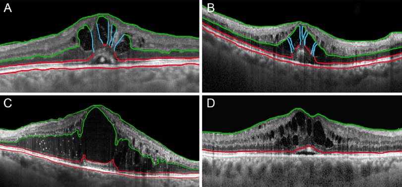
| Foveal eversion (FE) patterns in retinal vein occlusion: (A) is characterized by the presence of thick intraretinal vertical columns; (B) shows thinner intraretinal vertical columns, whereas (C) has no sign of columns in the context of intraretinal macular edema. RVO eyes with macular edema, showing a sign of foveal depression, are classified as “no FE” eyes. (D) Retinal vein occlusion. Photo: Arrigo, A., Aragona, E., Antropoli, A. et al. Foveal Eversion is Associated with High Persistence of Macular Edema and Visual Acuity Deterioration in Retinal Vein Occlusion. Ophthalmol Ther (2023). Click image to enlarge. |
A common complication of both central retinal vein occlusion (CRVO) and branch retinal vein occlusion (BRVO) is macular edema, which further causes reduced visual acuity. Foveal eversion describes a central fovea with a completely convex profile. In a previous study, a team of researchers based in Italy proposed that foveal eversion as a structural OCT biomarker is associated with a high prevalence of macular edema and poor outcome in diabetic retinopathy. Their most recent study determined that the presence of foveal eversion was associated with worse clinical course and outcome in CRVO and BRVO eyes.
The retrospective, observational case series included 168 eyes (168 patients) affected by CRVO and 116 eyes (116 patients) affected by BRVO. The researchers collected clinical and imaging data from CRVO and BRVO eyes affected by macular edema with a minimum follow-up of 12 months. On structural OCT, the team classified foveal eversion in cases that displayed it at baseline as pattern 1a, characterized by thick vertical intraretinal columns, pattern 1b, presenting thin vertical intraretinal lines, and pattern 2, showing no signs of vertical lines in the context of cystoid macular edema.
The study reported foveal eversion in 64 of 168 CRVO eyes (38%) and in 25 of 116 BRVO eyes (22%). Most of the eyes developed foveal eversion during follow-up. For CRVO eyes with foveal eversion, there were six eyes (9%) with pattern 1a, 17 eyes (26%) with pattern 1b and 41 eyes (65%) with pattern 2. Of those BRVO eyes with foveal eversion, there were eight eyes (32%) with pattern 1a and 1b and 17 eyes (68%) with pattern 2.
In both CRVO and BRVO eyes, the presence of foveal eversion was significantly associated with higher persistence of macular edema and worse outcome, with foveal eversion pattern 2 representing the most severe condition. Remarkably, foveal eversion patterns 1a and 1b were characterized by BCVA stability over follow-up, whereas pattern 2 showed significant BCVA worsening at the end of follow-up.
Foveal eversion characterized the minority of CRVO and BRVO eyes at baseline, rather than follow-up. In addition, the presence of peripheral capillary nonperfusion was slightly associated with worse clinical outcome and with the onset of foveal eversion.
“Although Müller cells are known to play a major role in the pathogenesis of diabetic retinopathy and diabetic macular edema, in this study we hypothesized their potential role also in the pathogenesis and clinical course of macular edema secondary to RVO,” the researchers wrote in their paper.
“Our study highlighted how foveal eversion, detected on structural OCT, can categorize more subtypes of CRVO and BRVO, characterized by different morphologies and functional outcomes,” they concluded. “From the perspective of customized treatment strategies and the adoption of artificial intelligence-based diagnostic approaches, foveal eversion might represent a useful metric to be included in the diagnostic workup of RVO.”
Arrigo A, Aragona E, Antropoli A, et al. Foveal eversion is associated with high persistence of macular edema and visual acuity deterioration in retinal vein occlusion. Ophthalmol Ther. June 9, 2023. [Epub ahead of print]. |
