A corneal specialist referred an 85-year-old white female to our military medical centers specialty contact lens clinic for a consultation. She had a long history of poor vision in both eyessince age 25, when she experienced an episode of Stevens-Johnson syndrome (SJS). Since then, she has been diagnosed with a sulfa drug allergy, primary open-angle glaucoma (POAG) and chronic ocular surface disease.
When she told us that she wanted to wear contact lenses, we were taken aback. How could someone with challenged dexterity and poor uncorrected acuity care for contact lenses properly?
But, she was motivated, and we thought that gas-permeable (GP) lenses might be able to not only improve her vision, but also to avoid aggravating any of her ocular conditions. Perhaps this patient could challengeand even exceedour expectations.
History
Bettys medical history was significant for an episode of SJS in 1946, which was induced by penicillin. Subsequently, she was found to be allergic to sulfa drugs.
A thorough assessment of her ocular history revealed documented eye exams by many optometrists and ophthalmologists from 1980 to 2006. Her history was also significant for diagnoses of POAG O.U., an epiretinal membrane O.S., long-standing corneal scarring (more O.S. than O.D.), and SJS with chronic ocular surface disease.

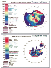
1. Topography revealed a highly irregular corneal surface O.D. (top), while readings were not even attainable O.S. due to excessive scar tissue.
Her corneal specialist was managing her with Muro 128 (sodium chloride, Bausch & Lomb), Patanol (olopatadine, Alcon), Restasis (cyclosporine, Allergan) and Tears Naturale (Alcon) for her chronic dry eye and ocular surface disease, and with Xalatan (latanoprost, Pfizer Ophthalmics) and Betoptic S (betaxolol 0.25% suspension, Alcon) for her glaucoma.
Betty had cataract surgery in her right eye in 1988 and in her left eye in 1990, as well as conjunctival grafts in both eyes in 1986. Surgery for trichiasis, entropion and tear duct implantation was performed in 1990. The tear duct implants were removed in 1994 because of chronic irritation. She was being followed routinely for her glaucoma as well as her ocular conditions caused by SJS.
Diagnostic Data
Her uncorrected visual acuities were 20/200+ (a pinhole acuity of 20/70) O.D. and 20/400 O.S. (no improvement with pinhole).
She said that she did not wear her glasses consistently for 60 years because they didnt help her subjectively. (She did not bring her spectacles with her.)

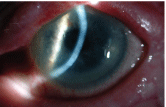
2. Slit lamp exam also found excessive scarring on the cornea O.S.
Her best-corrected visual acuity was 20/60 O.D. and 20/400 O.S., with a manifest refraction of +0.25D-6.00Dx115 O.D. and -2.25D-2.25Dx005 O.S. The manual keratometry readings were 45.00/44.50x095 O.D. and 44.50/42.74x095 O.S., with highly irregular mires.
Corneal topography showed a highly irregular corneal surface O.D., and we were unable to perform an accurate reading in the left eye because of the excessive corneal scar tissue (figure 1). Slit lamp examination showed central corneal scars on the left eye to a much greater degree than on the right (figure 2). Anterior chambers were wide open, irises were normal, and both eyes had clear posterior chamber intraocular implants. Her IOP measured 22mm Hg O.D. and 17mm Hg O.S. by applanation tonometry.
Betty seemed to vaguely recall an optometrist trying hard contact lenses on her eyes more than 30 years ago, but no lenses were ever dispensed. Since then, no other eye doctor discussed the possibility of rigid lenses with her.
With some misgivings about her age and ocular surface disease, we decided to give GP lenses a try. Over-refraction of both eyes with conventional spherical GP lenses resulted in a best-corrected visual acuity of 20/40+ O.D., but no improvement O.S. (it remained at 20/400). So, we ordered a Boston XO lens (base curve: 45.00, strength: -3.75D, diameter: 9.6mm) for her right eye and scheduled a follow-up for dispensing.
Follow-Up Exams
When Betty returned, we taught her how to insert and care for her lenses. As expected, both her dexterity and uncorrected vision made handling the lens challenging. However, she was eventually able to insert the lens with the aid of a magnifying mirror and could remove the lens with a DMV suction cup.
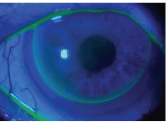
We allowed the lens to settle and the tear film to equalize for a few minutes. Over-refraction was +0.75D.S. and rendered 20/25-1 distance visual acuity O.D. Fluorescein evaluation showed a shallow central pool that extended toward the eight oclock position, which demonstrated the irregularity of the cornea (figure 3). The GP lens had good lid attachment, which helped it center well.
3. Fluorescein evaluation revealed a shallow central pool that demonstrated the irregularity of the cornea, but the lens centered well.
We recommended that Betty continue to use artificial tears and Restasis, and we recommended a conservative wearing schedule for the first week. We scheduled her for a one-week follow-up.

Eight days later, she was wearing the lens for nine hours each day with minimal complaints. Comfort was still an issue, as with all new GP lens wearers. The over-refraction of the right eye was then +0.50D.S. and dynamic visual acuity was 20/20-2 O.D. The fluorescein pattern remained unchanged. There were no epithelial erosions present, and there appeared to be plenty of tear film present for safe GP lens wear. We again observed insertion and removal procedures, and she was able to perform both fairly smoothly.
We scheduled two more follow-ups with her to check on her progress and ocular health. These subsequent follow-ups confirmed that she was determined to continue wearing her contact lens because it was the best vision she had experienced in 60 years, she said.
Nevertheless, complications still arise occasionally. For example, Betty struggled with lens dislocation twice in the past few months. Comfort is also still a minor issue if she wears her contact lens for more than five hours a day. Insertion and removal is still difficult for her, but she manages. We also remain uneasy about her ability to maintain adequate tear film. Still, Betty insists on keeping her contact lens, despite these lingering obstacles.
Discussion
Stevens-Johnson syndrome, also known as erythema multiforme major, is an acute mucocutaneous, vesiculobullous disease. SJS primarily occurs in young, healthy individuals, and it is more prevalent in females than males.1,2 The most common precipitating factors are a hypersensitivity reaction to drugs, such as sulfonamides, penicillin and salicylates, and infections caused by the herpes virus, adenovirus and mycoplasma pneumoniae.
Early signs include flu-like symptoms, followed by inflammation of the mucous membranes and a painful red rash that spreads and blisters. The basic lesion presents as an acute vasculitis that affects the skin and is concentrated on the hands, feet and mucous membranes.
SJS is an emergency medical condition that requires hospitalization. Treatment focuses first on eliminating the underlying cause. In most cases, it may be as simple as discontinuing a current medication. The second step is to control the symptoms and minimize any possible complications, such as inflammation and infection. Pain medication and antibiotics may be necessary, in addition to the replacement of bodily fluids and nutrients. Recovery can take weeks to several months, depending on the severity of the condition. The mortality rate of acute SJS ranges between 10% and 33%.2
SJS often results in severe ocular surface disease with poor visual prognosis. Symblepharon, adhesive occlusion of the lacrimal puncta, and corneal opacification with conjunctivalization are often observed in the chronic stages of the disease.1,2 Severe dry eye is another major concern in these patients, because it can lead to worsening ocular surface health.3
Treatment options for SJS patients concentrate on managing their ocular surface disease. Tear supplements and punctal occlusion can help manage tear deficiency.4 Topical corticosteroids and cyclosporine can help manage the inflammatory factors of chronic dry eyes.
Surgical options are also available. Corneal epithelial cells transplantation treats severe ocular surface disorders by reconstructing damaged corneal epithelial cells associated with SJS.3,5-7 Penetrating keratoplasty (PKP) and phototherapeutic keratectomy (PTK) can treat severe scar tissue secondary to ocular surface disease and improve vision.8
Scleral contact lenses for overnight wear have several benefits for SJS patients. These rigid, highly oxygen-permeable lenses protect the corneal surface from trichiasis, relieve dry eyes and improve vision.9,10
GP contact lenses offer similar advantages as scleral contact lenses, but no studies document their protective value against SJS. These rigid, oxygen-permeable contact lenses are used to mask corneal irregularity secondary to ocular surface disease, as was the case with this patient. The pre-corneal fluid reservoir can optically neutralize irregular corneal defects; so patients like Betty can potentially see much better when wearing GP lenses.
Betty was fortunate enough to survive a penicillin-induced SJS episode. Over the years, it has debilitated her vision and caused chronic dry eyes, corneal scarring and severe ocular surface disease.
We surmised that concerns about her age and long-standing ocular condition prevented optometrists and ophthalmologists from considering contact lenses. Indeed, we had to overcome those same concerns.
The rigid lens and adequate tear film masked most of the corneal irregularity (as expected) and caused no undue discomfort or corneal distress (as wed hoped). Acuity of 20/20- O.D. was a remarkable improvement when compared to the 20/200 O.D. she lived with for the last 60 years. With the visual benefit and subsequent motivation (both hers and ours), it was clearly worth the effort. Regular follow-up will be necessary to ensure continued ocular health. But, for now, this case shows that even at the age of 85, it is not too late to try.
Major Cang Q. Bui, O.D., is currently stationed at a forward deployed location in
1. Kanski JJ. Stevens-Johnson syndrome. In: Clinical Ophthalmology. A Systemic Approach. 3rd ed.
2. Kunimoto DY, Danitkar KD, Makar MS, et al. Stevens-Johnson syndrome. In: The Wills Eye Manual. 4th ed.
3. Nakamura T, Inatomi T, Sotozono C, et al. Transplantation of cultivated autologous oral mucosal epithelial cells in patients with severe ocular surface disorders. Br J Ophthalmol 2004 Oct;88(10):1280-4.
4. Kaido M, Goto E, Dogru M, Tsubota K. Punctal occlusion in the management of chronic Stevens-Johnson syndrome. Ophthalmology 2004 May;111(5):895-900.
5. Nakamura T, Ang LP, Rigby H, et al. The use of autologous serum in the development of corneal and oral epithelial equivalents in patients with Stevens-Johnson syndrome. Invest Ophthalmol Vis Sci 2006 Mar;47(3):909-16.
6. Nishida K, Yamato M, Hayashida Y, et al. Corneal reconstruction with tissue-engineered cell sheets composed of autologous oral mucosal epithelium.
7. Gomes JA,
8. Geerling G, Liu CS, Dart JK, et al. Sight and comfort: complex procedures in end-stage Stevens-Johnson syndrome. Eye 2003 Jan;17(1)89-91.
9. Fine P, Savrinski B, Millodot M. Contact lens management of a case of Stevens-Johnson syndrome: a case report. Optometry 2003 Oct;74(10):659-64.
10. Tappin MJ, Pullum KW, Buckley RJ. Scleral contact lenses for overnight wear in the management of ocular surface disorders. Eye 2001 Apr;15(pt 2):168-72.

