 |
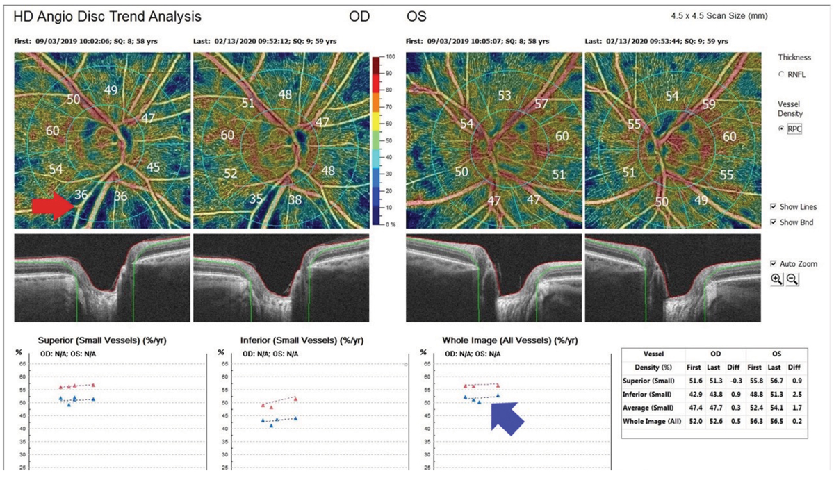
Reduced vessel density and microvascular dropout may also be the result of glaucomatous progression. Long-term changes in these parameters and their relationship to progression will require further investigation. Image above (not from the study) shows a nerve fiber layer defect (red arrow) and optic nerve perfusion improvement after treatment (blue arrow). Photo: Michael Cymbor, OD. Click image to enlarge. |
Use of OCT angiography (OCT-A) to evaluate ocular perfusion in the setting of glaucoma has been increasing. Though the modality hasn’t been widely adopted in clinical practice yet, experts expect that OCT-A will play a key role in the future of glaucoma diagnosis and follow-up. Swept-source OCT-A, a newer modality with projection artifact removal technique, is better suited to studying the microvasculature of the deep optic nerve head than spectral-domain OCT-A. Researchers assessed optic disc perfusion using swept-source OCT-A and standard-of-care visual field progression in primary open-angle glaucoma (POAG) eyes and found strong correlation with most parameters.
The researchers included 266 POAG eyes in the OCT-A and visual field evaluation, of which 30% showed visual field progression. In the progression group, there was a significantly higher proportion of disc hemorrhage, choroidal microvascular dropout and optic disc microvascular dropout but lower optic disc and parapapillary choroidal vessel densities compared with the non-progressors.
These parameters all demonstrated strong associations with one another. Based on those associations, the researchers developed models using different covariate combinations and found that younger age, presence of disc hemorrhage and lower optic disc vessel density were consistently associated with progression.
They concluded that optic disc hypoperfusion, represented as reduced optic disc vessel density along with peripapillary choroidal circulation as measured by OCT-A “showed association with visual field progression in POAG eyes. These OCT-A–based vascular parameters may contribute to monitoring of glaucomatous prognosis by facilitating prediction of glaucoma progression.”
Lee J, Sung K, Kim J, et al. Association between optic disc perfusion determined by swept-source optical coherence tomography angiography and visual field progression in glaucoma. J Glaucoma. [Epub ahead of print]. |
