 |
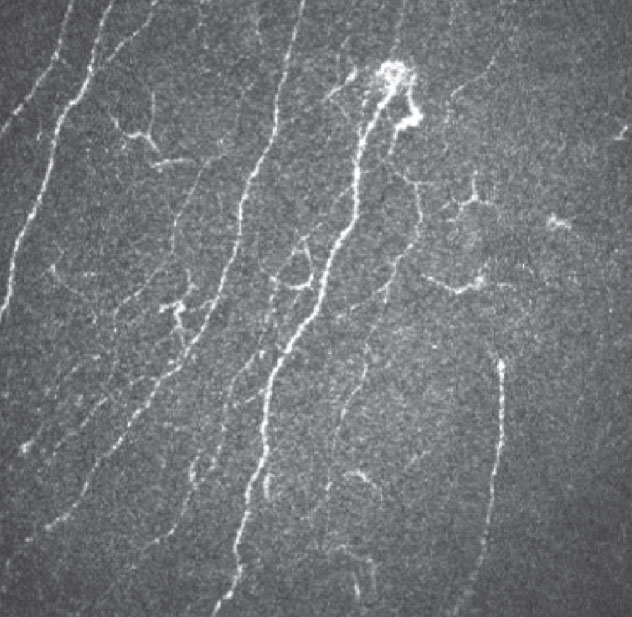
Individuals with autism are more likely to exhibit corneal structural alterations than those who are typically developing. Click image to enlarge. |
The relationship between connective tissue disorders and neurodevelopmental conditions has been established by previous reports, including higher rates of joint hypermobility. However, a new study is the first to examine the presence of corneal connective tissue structural alterations in individuals with autism spectrum disorder (ASD).
The study examined 30 ASD adults and 35 typically developing controls using in vivo confocal microscopy, specifically the K-structure in the Bowman's layer. K-structure grading was quantified into four levels, with grade 4 defined as “the mosaic K-structure is clearly seen in the image, and the background shows a significantly uneven wrinkled pattern. The keratocytes underneath the anterior stroma can be seen.” The ASD eyes received a significantly higher single-eye K-grading than the control eyes. Even when adjusted for inter-eye correlation and age, the ASD group’s K-gradings were still significantly higher than the control.
“To the best of our knowledge, the current study wrote the first to demonstrate connective tissue changes in the cornea in individuals with ASD,” said the study authors in the journal Autism Research. “Given that Bowman’s layers are mostly composed of collagen fibers I and V, our finding provides novel and preliminary evidence supporting the possible presence of connective tissue abnormalities in ASD individuals.”
In the study, K-grading didn’t correlate with autistic severity, but the authors found it to be positively associated with low visual sensory registration in the controls and negatively associated with visual sensory seeking in ASD. “In other words, ASD individuals seem to be less responsive to certain sensory stimulation and are less likely to seek sensation stimulation,” stated the authors. “Based on our findings, the presence of corneal connective tissue abnormalities in ASD individuals might partially explain the lower visual sensory registration and reduced visual sensation seeking observed in ASD.”
Larger scale studies of both males and females are needed, as the authors cited limitations in their study, including the fact that only male ASD adults were analyzed, despite the fact that females are reported to have a higher prevalence of generalized joint hypermobility and other connective tissue symptoms.
In summary, the researchers wrote, “Our in vivo confocal microscopy results could serve as a foundation for subsequent histological analyses and investigation of the possible relationship between connective tissue abnormalities and neurodevelopmental disorders.”
Chien YL, Wu PY, Wu JH, Huang WL, Hsiao CC, Hsieh YT, Cheng T, Gau SS, Chen WL. Corneal structural alterations in autism spectrum disorder: An in vivo confocal microscopy study. Autism Res. December 5, 2023. |
