
On examination, best-corrected visual acuity was 20/40 O.U. Confrontation fields were full to careful finger counting O.U. Pupils were equally round and reactive with no afferent pupillary defect. Motility testing and color vision were normal in each eye. The anterior segment was unremarkable.
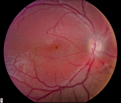
Dilated fundus exam showed small cups with good rim coloration and perfusion O.U. There was a marked absence of the foveal light reflex. Also, there were peculiar changes as shown in figures 1 to 2 and as shown enlarged in figures 3 and 4. We ordered optical coherence tomography (OCT), as shown in figures 5 and 6.
 |
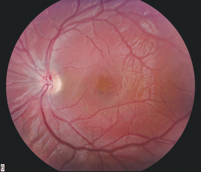 |
| 1, 2. Color fundus photos (O.D. left, O.S. right) showing suspicious changes in the macula. | |
 |
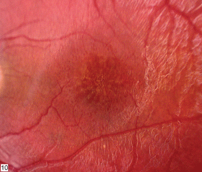 |
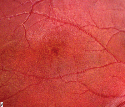
| 3, 4. These 2x fundus photos of the macula (O.D. left, O.S. right) show more detail of the macular lesions. The maculae are best described as having a petaloid pattern appearance, similar to the appearance of cystoid macular edema. | |
 |
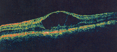 |
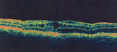
| 5, 6. (O.D. left, O.S. right) shows the corresponding macular lesions. Note the empty clear spaces within the sensory retina at the inner nuclear layer. What do these represent? |
Take the Retina Quiz
1. What do the changes in the macula represent?
a. Beaten metal appearance.
b. Cystoid macular edema.
c. Macular hypoplasia.
d. Macular schisis.
2. What is the correct interpretation of the OCT?
a. Cystoid macular edema.
b. Hypoplastic macula.
c. Internal limiting drape.
d. Macular schisis.
3. What other findings would you expect to see in this patient?
a. Vitreous cells.
b. Posterior subcapsular cataract.
c. Peripheral findings.
d. Quiet choroid.
4. What is the correct diagnosis for this patient?
a. Pars-planitis.
b. X-linked juvenile retinal schisis.
c. Stargardts macular dystrophy.
d. Cone dystrophy.
(For answers to this months Retina Quiz, please see below.)
Discussion
Our patient presented with bilateral, symmetric macular lesions that indicate a dystrophy. The family historythe brother whose eye problem necessitated surgeryalso suggests a dystrophy.
The maculae are best described as having a petaloid pattern appearance, similar to the appearance of cystoid macular edema. However, that is not what our patient has. We did not order a fluorescein angiogram, but had we done so, it likely would have been normal.
Based on the clinical appearance, the OCT findings and the family history, we diagnosed our patient as having X-linked juvenile retinoschisis.
X-linked juvenile retinoschisis is almost exclusively seen in males.1 The macular lesions are typically present at or shortly after birth.
However, the condition usually is not recognized until after the child starts school. It is usually diagnosed before age 15. Visual acuity ranges from 20/40 to 20/200, and slowly declines.
The schisis disappears later in life, leaving an atrophic macular scar.
About 50% of patients with X-linked juvenile retinoschisis have a peripheral schisis that is usually bilateral and located in the inferotemporal portion of the macula.1 Peripheral schisis represents a split between the inner nuclear and outer plexiform layers of the retina. Both inner and outer wall holes can be present.
About 10% to 25% of patients who have peripheral schisis also have a combined retinal detachment.1 In rare situations, vitreous veils, sheathing of the retinal vessels and vitreous hemorrhage can also develop.
Our patients older brother developed a combined retinal detachment that necessitated surgery.
A foveal schisis represents superficially localized cysts that are arranged in a stellate, or radial, pattern. This develops from a splitting of the nerve fiber layer.
On OCT, these appear as empty clear spaces within the sensory retina at the inner nuclear layer. Distinct walls separate the cystic spaces.
The diagnosis of retinoschisis usually is based on clinical findings and a good case history. OCT, fluorescein angiography and electrophysiology testing can help confirm suspicions.
An electroretinogram, or ERG, will show reduced scotopic and photopic b-wave amplitudes, which indicates X-linked retinoschisis. This is more apparent in the dark-adapted state than light, and it reflects functional impairment of the inner retinal layers.
X-linked juvenile schisis is caused by mutations in the RS1 gene. This gene encodes the discoidin domain protein retinoschisin, which is secreted by photoreceptors and bipolar cells.2
Our patient had macular schisis but no peripheral retinal schisis. He currently has no visual symptoms. Although his visual acuity is reduced, he reported no functional impairment in school or while performing any other activities.
We also examined the patients younger brother, but he showed no signs of the disease. We continue to monitor the family closely.
Retina Quiz Answers: 1) d; 2) d; 3) c; 4) b.
1. Edwards AO, Robertson JE. Hereditary vitreoretinal degenerations. In: Ryan SJ, ed.. Retina. 4th ed. St. Louis, Mosby. 2001:519-37.
