 A 61-year-old white female with a history of colon adenocarcinoma was referred for evaluation of “multiple white lesions” in both eyes. She had no significant ocular history, but was concerned about the possibility of metastasis from her history of colon cancer. At the time, she was undergoing chemotherapy and Avastin (bevacizu-mab, Genentech/Roche) therapy.
On examination, her best-corrected visual acuity measured 20/20 O.U. Confrontation fields were full to careful finger counting O.U. Extraocular motilities were full O.U. Her pupils were equal, round and reactive to light, without evidence of afferent defect.
The anterior segment evaluation was unremarkable. Her intraocular pressure measured 16mm Hg O.U. Dilated fundus exam revealed small cups with good rim coloration and perfusion in both eyes.
The vessels and maculae were unremarkable O.U. The peripheral retina examination revealed the changes seen in figures 1 and 2. Additionally, we performed fluorescein angiography (FA) imaging (figures 3 and 4).
Take the Retina Quiz
1. Which additional tests would be most helpful in establishing a diagnosis?
a. Standardized ultrasound.
b. Optical coherence tomography.
c. MRI.
d. Visual fields.
2. What is the correct diagnosis in this case?
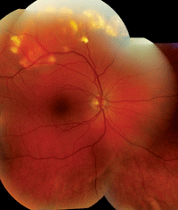
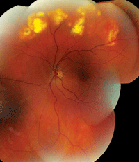
1, 2. The peripheral retinal images show peculiar white lesions in both eyes (O.D. left, O.S. right). Are these a cause for concern?
a. Metastatic carcinoma to the choroid.
b. Coats’ disease.
c. Choroidal osteoma.
d. Sclerochoroidal calcification.
3. How should this patient be managed?
a. Plaque radiotherapy.
b. Systemic chemotherapy.
c. Observation.
d. Enucleation.
4. What’s the prognosis for this patient?
a. Excellent.
b. Guarded.
c. Poor.
d. Too early to tell.
For answers, see below.
Discussion
Our patient has sclerochoroidal calcification––a rare condition characterized by the presence of irregular, yellow-white subretinal placoid lesions usually seen in the superotemporal mid-periphery of the fundus. The presentation commonly is misdiagnosed as a malignant tumor.
Sclerochoroidal calcification typically is idiopathic and often is documented as an incidental finding in elderly patients (although it can be associated with hyperparathyroidism, vitamin D intoxication, sarcoidosis, hypophosphatemia or chronic renal failure).1,2 The calcifications are believed to be deposited at the insertion sites of the extraocular oblique muscles.1 Nonetheless, the condition remains a poorly recognized and understood entity.
Because sclerochoroidal calcification exhibits a very characteristic clinical appearance and anatomical location, the diagnosis should be easy to make––so long as the clinician is aware of the condition and maintains a heightened level of suspicion. Additionally, FA and ultrasound can help confirm the diagnosis.
In the early phases of the angiogram, FA will reveal autofluorescence of the lesions with the red-free filter and mild-to-moderate hypofluorescence.
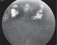
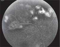
3, 4. Late-phase fluorescein angiography shows staining and hyperfluorescence of
the lesions (O.D. top, O.S. bottom).
In the later phases, the lesions will show increased hyperfluorescence as well as persistent late staining and mottling around the lesion that corresponds to areas of RPE atrophy. This is exactly what occurred in our patient.
The ultrasound finding—also typical of sclerochoroidal calcification—shows high reflectivity of the lesion’s anterior surface as well as intense echodense placoid lesions at the scleral-choroidal interface with orbital shadowing.2 According to one study, sclerochoroidal calcification chiefly involves the choroid; however, the sclera also may be involved.2 In this case, the lesions had a maximum thickness of 2.2mm.
Given their creamy white appearance, sclerochoroidal calcifications often are mistaken for choroidal osteomas––a benign ossifying tumor comprised of mature bone that forms within the choroid.
Osteomas commonly are located immediately adjacent to or around the optic nerve and can extend to the macula. They are yellow-white to orange-red in color (similar to a sclerochoroidal calcification), and will exhibit high reflectivity and posterior shadowing on ultrasound due to their bony configurations. In fact, sclerochoroidal calcifications initially were believed to be choroidal osteomas that presented in older patients. Subsequent histological studies revealed the identifiable differences between these lesions.
Lastly, choroidal metastasis must be ruled out, because these lesions also tend to exhibit a creamy white appearance and often present bilaterally. With our patient’s history of colon cancer, this was a legitimate concern. Fortunately, cancerous tumors are easily differentiated on ultrasound. Choroidal metastasis shows medium to high reflectivity, but lacks the shadowing of sclerochoroidal calcifications.
We explained the findings to our patient, and she was relieved to learn that the retinal changes were benign and unrelated to her history of colon cancer.
At both three- and six-month follow-up, we detected no signs of progression. We will continue to follow her every six months for a year, then annually thereafter.
Thanks to former Bascom Palmer optometry resident Melanie Denton, O.D., of Burnsville, N.C., for contributing this case.
1. Cooke CA, McAvoy C, Best R. Idiopathic sclerochoroidal calcification. Br J Ophthalmol. 2003 Feb;87(2):245-6.
2. Honavar SG, Shields CL, Demirci H, et al. Sclerochoroidal calcification: clinical manifestations and systemic associations. Arch Ophthalmol. 2001 Jun;119(6):833-40.
Answers
1. a
2. d
3. c
4. a
