 |
Q: A healthy, long-time soft lens patient has a significant fibro-vascular pannus (FVP) superiorly, epithelial insult, mild limbal hy- peremia and a relatively quiet bulbar conjunctiva. A corneal specialist thinks this might be a stem cell deficiency. Can this happen secondary to soft lens wear? If so, whats the best treatment?
A: A stem cell is a cell that can develop into many different cell types in the body.1 It serves as a repair system for the body and thus, can theoretically divide without limit to restore other cells.1 When a stem cell divides, each new cell can either remain a stem cell or become another type of cell with a more specific function, such as a brain cell.1
So, can stem cell deficiency happen secondary to soft lens wear? Any contact lens that rides on or touches the limbus can contribute to stem cell deficiency, says optometrist Christine Sindt, director of the contact lens service at the University of Iowa, in Iowa City.
 |
|
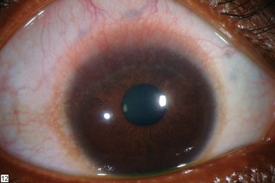
Initial presentation of a different patient diagnosed with stem cell deficiency. |
In certain pathologic conditions, such as contact lens-related trauma or inflammation, stem cells may be partially depleted. So, the patient has fewer signs and symptoms of stem cell deficiency, and the conjunctiva often looks uninvolved.
But in cases of severe chemical burns or autoimmune diseases, such as Stevens-Johnson syndrome (SJS) and ocular pemphigoid, stem cells can be completely destroyed. This results in varying degrees of stem cell deficiency with such characteristic features as corneal stromal scarring and severe staining.
In more advanced cases of contact-lens related stem cell deficiency, however, the cornea has some vascularization and an irregular and unstable corneal epithelium. This ultimately leads to conjunctivalization and corneal stromal involvement if left unchecked.
A newly devised classification system uses the extent of the patients limbal stem cell deficiency and the appearance of the conjunctiva to classify the disease. This allows for treatments specifically tailored to each patient.2 (See the referenced article for further information.)
So whats the best course of action for this patient?
Discontinue lens wear. If the patient recovers rapidly (within 24 to 72 hours), the ocular surface compromise is more likely due to a solution hypersensitivity or hy-poxia. In this case, place the patient back in contact lens wear following resolution, and monitor her periodically over the next several weeks, Dr. Ryan says. If the condition progresses, be suspicious that this represents true stem cell deficiency related to some other mechanism.
Generally, contact lens keratopathy results from mechanical and/or contact lens solution issues. So, it is a less severe form of stem cell deficiency and does not require significant intervention such as surgery.
Prescribe non-preserved topical lubricants. Have the patient use non-preserved artificial tears, q.i.d. to q1h depending on the patients signs and symptoms, for the long-run, if it proves helpful. If the process stabilizes and the patient is comfortable, just follow her, says Barry Weissman, O.D., of the Jules Stein Eye Institute in Los Angeles. Follow-up intervals depend on the patients signs and symptoms. If, however, the disease progresses, consider surgery.
Refer the patient for surgery. The signs and symptoms of a severe form of stem cell deficiency include persistent epithelial defects, conjunctival injection, chronic irritation, neovascular pannus and, eventually, stromal scarring. There can be an abnormal stem cell population or a decreased number of stem cells, but usually both.
If this is the case, this patient has two advanced surgical options: limbal cell transplantation and amniotic membrane transplantation. In limbal cell transplantation, the surgeon removes limbal stem cells from the patients healthy eye (autograft) or from the normal eye of a cadaver or living relative (allograft) and transplants that section to the stem cell-deficient eye.3
In amniotic membrane transplantation, the surgeon removes the inner amniotic membrane from fetal membranes, which consists of a single layer of amnion cells.4 The surgeon then overlays the entire corneal surface, including the limbus, with the amniotic graft, which acts as a biological contact lens.4
While it is unclear whether amniotic membrane fosters limbal cell proliferation, studies have shown that it does foster epithelialization of the corneal surface.4 This technique can be used in conjunction with limbal stem cell transplantation and is most beneficial in patients who have total stem cell deficiency.4 Still, Dr. Weissman is on the fence regarding this procedure, as there have been questions about the efficacy of it in this disease, he says.
1. Stem Cell Basics. National Institutes of Health (NIH). http://stemcells.nih.gov/info/basics. (Accessed February 17 2006).
2. Simon-Hidalgo A. New classification system to assist in diagnosis and treatment of limbal stem cell disease. www.escrs.org/eurotimes/April2003/newclassification.asp. (Accessed February 17 2006).
3. Karpecki PM. Improved vision stems from cells. Rev Optom 2004 Jul;141(827):88-9.
4. Schwan BL. Human amniotic membrane transplantation for the treatment of ocular surface disease. Duval County Medical Society. Northeast Florida Medicine Journal. www.dcmsonline.org/jaxmedicine/2002journals /augsept2002/amniotic.htm. (Accessed February 15 2006).
