 |
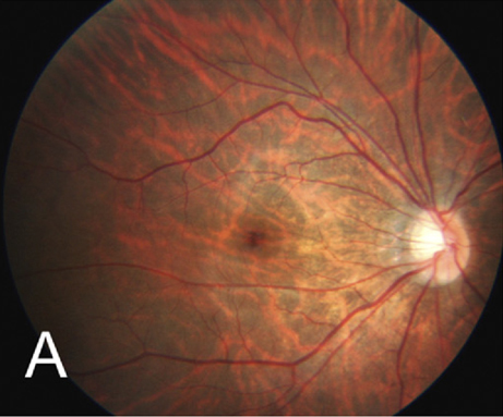
| Fundus tessellation is a subtype of myopic maculopathy characterized by enlarged choroidal vessels around the fovea with thinning of the retinal epithelium and choriocapillaris. Photo: Hayashi K, Ohno-Matsui K, Shimada N, et al. Ophthalmology. 2010;117:1595-161. Click image to enlarge. |
Eyes with high myopia are at risk for a number of retinal complications. Fundus tessellations, one of the initial stages of myopic maculopathy, may indicate other myopic changes down the line. Recently, researchers conducted a systematic review and meta-analysis to better understand fundus tessellations, since the current body of literature, though vast, varies significantly in content and research direction, and includes some conflicting reports. Their findings, published recently in Ophthalmology and Therapy, showed that prevalence of fundus tessellations varies based on population characteristics and has several key differences from a normal fundus.
Their systematic review included 23 articles (3,053 eyes) whose data were evaluated based to the Newcastle-Ottawa Scale and the Healthcare Research and Quality criteria. The meta-analysis aimed at comparing tessellated and normal fundus.
The researchers reported that the prevalence of fundus tessellations ranged between 43% and 94.35%. Severity was significantly associated with older age, male sex, lower body weight index, longer axial length, larger peripapillary atrophy, thinner choroid, thinner sclera and larger corneal radius of curvature that the researchers noted suggested “a potential progression pattern.”
They also found that fundus tessellations were predominantly seen in the macular and peripapillary regions. Tessellated fundus was associated with older age, longer axial length and lower spherical equivalent vs. normal fundus in the meta-analysis. The researchers reported that there was no significant difference in the proportion of males among those with tessellated and normal fundus.
They concluded that these findings could bolster research for improved diagnostic precision and patient care. However, they pointed out in their Ophthalmology and Therapy paper that “the lack of a standardized classification hampers comparability and generalizability of research across studies. Furthermore, the diverse research objectives regarding fundus tessellations result in variations in the ages, genders, and refractive states of the study subjects. This complexity makes it difficult to conduct subsequent comparisons and summaries.”
They wrote that a standardized classification system and large-scale databases are needed. “Such a system would enhance the consistency and accuracy of assessments, enabling better comparisons between studies and an improved understanding of the condition’s progression and associated factors.”
Chen X, He H, Xu J, et al. Clinical features of fundus tessellation and its relationship with myopia: a systematic review and meta-analysis. Ophthalmol Ther. September 21, 2023. [Epub ahead of print]. |
