
History
A 32-year-old black female presented for examination with a chief complaint of bilateral temporal eyelid edema along with tearing of two weeks duration. Earlier in the day, another optometrist saw her and referred her to our clinic. She had self-medicated with cold compresses and Benadryl (diphenhydramine, Pfizer) with no relief. Yet she reported no allergies. Her ocular and systemic history was unremarkable.
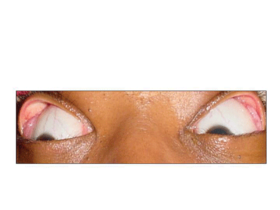
Best-corrected visual acuity at distance and near measured 20/20 O.D. and 20/20 O.S. Pupils were round and reactive to light with no afferent defect. Extraocular muscles were full and smooth. Confrontation visual fields were normal. Anterior segment evaluation was unremarkable. A more detailed external examination revealed prominent accessory lacrimal glands. The photograph demonstrates the clinical findings.
Dilated fundus examination revealed healthy optic nerves in both eyes with normal cup-to-disc ratios and clear maculae with no retinal holes, tears or detachments.
Your Diagnosis
How would you approach this case? Does this patient require additional tests? What is your diagnosis? How would you manage this patient? What is the likely prognosis?
Discussion
Additional tests center around identifying the underlying cause for the glandular swelling. Possible underlying causes include infectious or infiltrative diseases such as sarcoidosis, rheumatoid arthritis, lupus, Lyme disease, orbital pseudotumor, syphilis, tuberculosis and Wegeners granulomatosis. The full laboratory workup should include: complete blood count (CBC) with differential and platelets, erythrocyte sedimentation rate (ESR), antinuclear antibody (ANA), antineutrophilic cytoplasmic antibodies (ANCA), fluorescent treponemal antibody absorption test (FTA-ABS), rapid plasma reagin (RPR), purified protein derivative (PPD), chest X-ray, angiotensin converting enzyme (ACE), serum lysozyme, Lyme immunofluorescent antibody titer, enzyme-linked immunosorbent assay for IgM and IgG, and HLA-B27.
The diagnosis in this case is bilateral sarcoidic infiltration of the lacrimal glands.
Sarcoidosis is a multisystem disease that predominantly affects the lungs, liver and central nervous system.1-7 The etiology of sarcoid is unclear.1-7 The pathologic findings are believed to be the result of an immunologic problem, in which unknown antigens
initiate a systemwide inflammation and infiltration.1-7 Lymphocytes and mononuclear phagocytes are attracted to the affected area and become important in the formation of noncaseating granulomas, the histologic hallmark of the disease.4
Clinical presentation depends on the organ system or tissues affected.1-4 A majority of patients demonstrate pulmonary involvement in the form of alveolitis, followed by granulomatous infiltration and fibrosis.1-5 Neurologic manifestations commonly involve the cranial nerves and meninges. Facial nerve palsy is the most common sign of neurosarcoidosis. The liver, spleen and heart may also become infiltrated.5
Ocular manifestations can be among the first signs of the disease and may involve all structures of the eye.1-7 A large number of patients develop chronic granulomatous iridocyclitis with mutton fat keratic precipitates, Koeppe and Busacca iris nodules, and snowballs in the anterior vitreous. Posterior synechiae may lead to iris bombe and angle closure glaucoma. Keratoconjunctivitis sicca may develop secondary to lacrimal gland infiltration.1-4
Other issues of this condition include conjunctival nodules leading to progressive cicatricial conjunctivitis and posterior segment findings such as periphlebitis or chorioretinitis. Cystoid macular edema, retinal neovascularization, disc edema and optic nerve granulomas can also occur.1-4
Sarcoidosis can be diagnosed with serum lysozyme and angiotensin converting enzyme studies in conjunction with chest radiography. Other useful testing includes a limited gallium scan of the head and neck or a biopsy of suspicious skin, conjunctival or lacrimal gland lesions.6,7
The mainstay of sarcoid treatment is oral corticosteroids. For systemic therapy, a two-week loading dose of 60 to 80mgs q.d. p.o., followed by a six week taper, and then a minimum six-month maintenance dose (which may range from 5 to 20mgs q.d. p.o. depending on the severity) is usually required. The treatment of ocular sarcoidosis varies depending on the tissues involved and the extent of the damage.1-5
The mortality rate in sarcoidosis is primarily due to the complications of pulmonary insufficiency and cardiac involvement.6,7
1. Belfer MH, Stevens RW. Sarcoidosis. Sarcoidosis: a primary care review. Am Fam Physician 1998 Dec;58(9):2041-50, 2055-6.
2. Hunninghake GW, Gilbert S, Pueringer R, et al. Outcome of the treatment for sarcoidosis. Am J Respir Crit Care Med 1994 Apr;149(4 Pt 1):893-8.
3. Paramothayan S, Jones PW. Corticosteroid therapy in pulmonary sarcoidosis: a systematic review. JAMA 2002 Mar 13;287(10):1301-7.
4. Rothova A. Ocular involvement in sarcoidosis. Br J Ophthalmol 2000 Jan;84(1):110-6.
5. Thomas KW, Hunninghake GW. Sarcoidosis. JAMA 2003 Jun 25;289(24):3300-3.
6. Yanardag H, Pamuk ON. Lacrimal gland involvement in sarcoidosis. The clinical features of 9 patients. Swiss Med Wkly 2003 Jul 12;133(27-28):388-91.
7. Yanoff M, Fine BS. Granulomatous Inflammation. In: Yanoff M, Fine BS. Ocular Pathology. St. Louis, Mo.: Mosby; 2002:96-99.
Thanks to Lori Ives, O.D., for contributing this case.
