 |
Over the past five years, this column has covered many topics related to lenses and prisms, but it has not yet discussed yoked prisms in detail. As we all learned in optometry school, a prism shifts light, and the person shifts their eye in response. Specifically, yoked prisms shift images the same direction and amount in both eyes.
If you stick with the basic understanding of prisms as chiefly ray shifters (1.00cm for every 1.00m traveled per 1.00D of prism), you will miss their true power. Besides their ability to shift light and eyes, yoked prisms initiate spatial shifts, or shifts in a person’s center of gravity, causing an observed postural change, which triggers a shift and/or rotation in the pelvis.1 These changes have the potential to alter behavior and attention.
In the field of neuro-optometric rehabilitation, we use horizontal yoked prisms in the treatment of cases of homonymous hemianopsia, midline shift, neglect and sometimes unilateral spatial inattention. In patients with nystagmus who have a null point that requires a head turn, yoked prisms sometimes allow for the patient to maintain a straighter head position, alleviating future spinal issues.
Vertically oriented yoked prisms are used most often to improve behavior and attention. While the exact mechanisms are not fully understood, we know these prisms transform space beyond the shifts in the chief ray in the direction of the apex of the prism. Other spatial transformations are complex but understood.1 We find that patients on the autism spectrum, as well as those with attention difficulties, often respond positively to vertical yoked prisms.
The goal with prism is to improve and enhance looking behavior and to extend how long a patient is able to pay attention. This column focuses on the use of vertically oriented yoked prisms and includes two stories of success.
 |
|
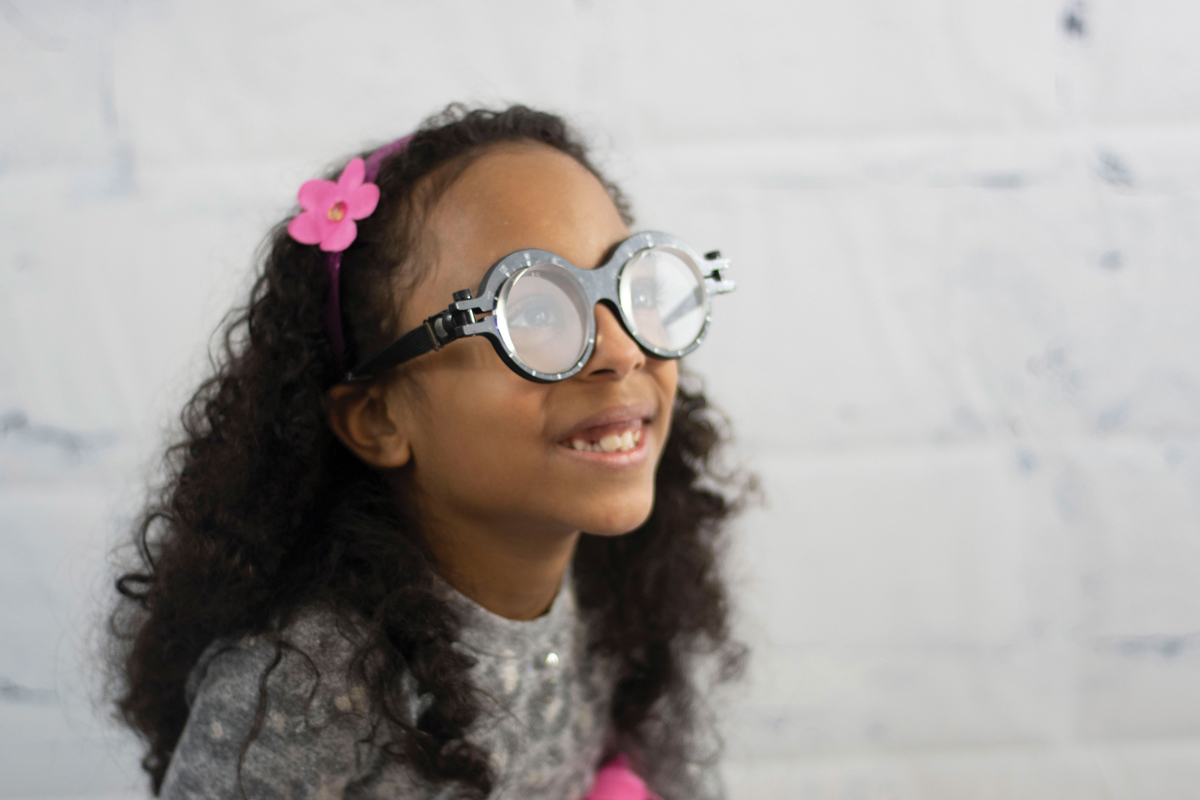
This patient is wearing base-up yoked prism. Click image to enlarge. |
Case One
A six-year-old girl had an eye exam several weeks prior, but her father was concerned that she was still moving her body instead of her eyes while writing. The patient’s medical history included ADHD and autism. She uses both Focalin (dexmethylphenidate, Novartis) and guanfacine. She was born on time and has been meeting all developmental milestones but is mildly delayed. She is not currently reading and receives speech and occupational therapy several times per week. Her father described her as “very clumsy” and said she bumps into things a lot at home.
The patient was able to see 20/15 OD, OS and OU, had 20 seconds of stereo and was orthophoric at distance and near. Retinoscopy was +0.50 -0.50x090 OU. The monocular estimated method response through plano was +0.50 with fluctuations. Through +0.50, the response was plano with fewer fluctuations. A test of gross eye movement demonstrated significant issues on both pursuits and saccades, showing significant body overflow and head movement. When we were able to get her to sit down, the patient was fidgety in the chair, and she was jumpy otherwise.
We decided to trial 2.00D of base-up yoked prism. Interacting with the hanging Marsden ball, the patient’s attention was more focused with the yoked prism and she was better able to catch the ball. Her father commented that he had never seen his daughter so focused and with such good hand-eye coordination.
We prescribed +0.25D of sphere with the 2.00D of base-up yoked prism OU and initiated a course of vision therapy focusing on eye and body movements. The patient is currently on her 10th session and progressing wonderfully.
 |
|
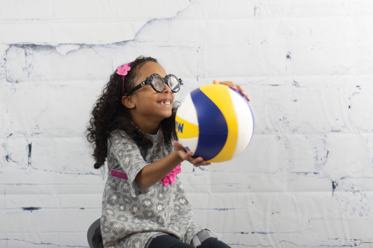
The patient, while wearing base-up yoked prisms, catches a ball during a visual performance assessment. Click image to enlarge. |
Case Two
A minimally verbal four-year-old boy with autism was referred by his occupational therapist for a visual evaluation due to difficulty with visual attention to tabletop tasks and moving targets, as well as visual motor integration concerns. He receives occupational therapy services for sensory processing, visual motor integration, fine motor skills, bilateral integration, gross motor skills and difficulty with daily activities. He also receives speech therapy.
The patient had a limited attention span, so only the basic examination components and some modified visual perceptual tests were performed. He was able to see 20/20 OU at distance and 20/25- OU at near with Lea symbols. He did not like either eye being covered, so monocular acuities were not evaluated. His eye movements showed no restrictions but were jerky in nature and consisted of small microsaccades instead of a smooth saccade or pursuit movement.
His visual attention to our target was very limited (about one to two seconds at a time). His cover test revealed orthophoria at distance and near, and his near point of convergence was to the nose on two attempts; after that, he lost interest and attention. His distance retinoscopy was plano OD and OS, and his just-look retinoscopy showed bright, equal reflexes with a near retinoscopy finding of +0.50D of sphere OD and OS for the brightest, most stable reflex.
We chose to trial a higher amount of prism than usual to see if there was a positive response, since the patient’s visual attention was so fleeting. He wore 5.00D of base-up yoked prism and then 5.00D of base-down yoked prism while walking and interacting with the Marsden ball and other various objects around him. He walked in a straighter line and was more attentive to his surroundings with the base-up yoked prism; however, his attention was still limited. With the base-down yoked prism, he had an immediate negative response, was very anxious and ripped the glasses off.
At the following visit, we attempted testing again but did not get the response we had hoped for. During basic perceptual testing, the patient looked at each object for one to two seconds before looking away and then had to be redirected to where the object was on the table several times before he would visually attend.
We trialed a lower amount of prism this time (2.00D OU), and the response was instantaneous. The patient’s attention improved and he completed tasks that were previously challenging. We prescribed +0.25D of sphere with the 2.00D of base-up yoked prism OU.
The patient was re-evaluated two months later. His mom noted that he had been wearing his glasses at school, during occupational therapy and occasionally on the weekends at home when doing near tasks. He was able to perform several perceptual tests that he previously could not complete. Since he was already involved in so many other therapies, we decided to hold off on vision therapy. We monitor his progress every three to six months.
Takeaways
In patients with ADHD or autism who may benefit from yoked prism, we often try smaller amounts (1.00D to 5.00D) of base-up and base-down yoked prism per eye. The goal is to provide the smallest amount of yoked prism to create a positive change in awareness and a subsequent response. Higher amounts can also be trialed, but the goal remains the same. Typically, 1.00D to 3.00D of prism per eye is prescribed.
While seeing these patients can be challenging, you have the skill and knowledge to succeed. Not only can you improve the clarity of sight, but you can also enhance vision and encourage development of the visual process through the use of vertically oriented yoked prism.
Dr. Taub is a professor, chief of the Vision Therapy and Rehabilitation service and co-supervisor of the Vision Therapy and Pediatrics residency at Southern College of Optometry (SCO) in Memphis. He specializes in vision therapy, pediatrics and brain injury. Dr. Harris is also a professor at SCO. Previously, he was in private practice in Baltimore for 30 years. His interests are in behavioral vision care, vision therapy, pediatrics, brain injury and electrodiagnostics. They have no financial interests to disclose.
Dr. Groce is an assistant professor and the residency supervisor for the Vision Rehabilitation and Brain Injury Residency at SCO. She also sees patients in the Eye Center’s pediatrics, teen and vision therapy clinics. She has no financial interests to disclose.
1. Harris J. The use of lenses to improve quality of life following brain injury. In: Suter PS, Harvey LH, eds. Vision Rehabilitation: Multidisciplinary Care of the Patient Following Brain Injury. Oxfordshire, England: Routledge; 2011. |
