 |
|
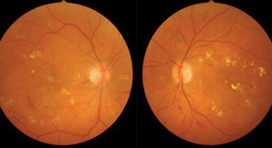
Hyperreflective foci may indicate macular edema progression and help guide treatment in affected patients. Photo: A. Paul Chous, OD. |
Macular edema is usually controlled with anti-VEGF or anti-inflammatory agents, but patient response to treatment is often inconsistent. To better understand different causes of macular edema and choose the most suitable treatment option, researchers recently examined the macular microstructural features of these patients using spectral domain OCT images. They found that hyperreflective foci may indicate disease prognosis and may be an important characteristic to consider when deciding on a treatment regimen.
The retrospective study included the following patient groups:
101 eyes of 86 patients with diabetic macular edema (DME)
51 eyes of 51 patients with macular edema secondary to branch retinal vein occlusion (BRVO-ME)
59 eyes of 58 patients with macular edema secondary to central retinal vein occlusion (CRVO-ME)
26 eyes of 22 patients with uveitic macular edema
The researchers reported that the number of hyperreflective foci, the frequency of hard exudates and the enhanced internal reflectivity of the cysts were significantly different among the four groups. The DME group had significantly higher numbers of hyperreflective foci than the other groups. The frequency of hard exudates and enhanced internal reflectivity of cysts in the DME group was also significantly higher than in the BRVO-ME and CRVO-ME groups. The researchers added that in the DME group, the number of hyperreflective foci in patients with hard exudates was significantly higher than in patients without.
They concluded that hyperreflective foci were more frequent in DME patients and that the occurrence of hyperreflective foci was correlated with the frequency of hard exudates. “We speculated the hyperreflective foci in DME patients represent the deposition of large molecular exudates such as protein and lipids in retinal tissue, which may be the precursor to hard exudates,” the researchers wrote in their paper. “Hyperreflective foci in DME patients may be an important sign of blood-retinal barrier destruction.”
Zhu R, Xiao S, Zhang W, et al. Comparison of hyperreflective foci in macular edema secondary to multiple etiologies with spectral-domain optical coherence tomography: an observational study. BMC Ophthalmol. 2022;22:352. |
