 |
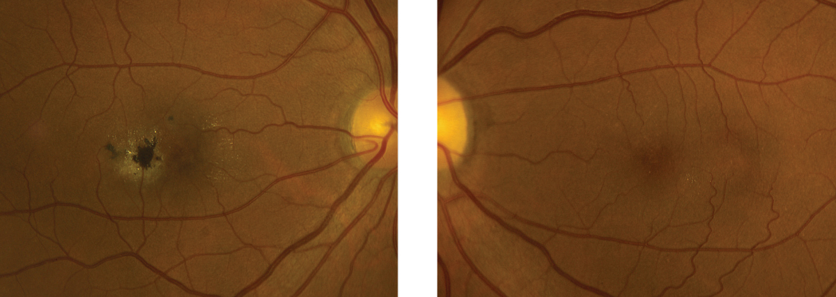
| Together, microperimetry and multifocal electroretinography can be used as tools to accurately assess the macula. Photo: Eric Dillinger, OD. Click to enlarge. |
When it comes to diagnosing type 2 macular telangiectasia (MacTel), new research suggests both microperimetry and multifocal electroretinography may work well together as valuable, noninvasive tools for functional assessment of the macula. In tandem, both methods showed a distinct pattern of localized macular dysfunction and provided complementary insight into inner and outer retinal changes, the researchers explained.
“The results provide further evidence to confirm the inner retina as the primary site of dysfunction, furthering our understanding of the pathophysiology of the disease, and may help identify biomarkers to monitor therapeutic clinical trials,” the authors wrote in their paper.
The study included 35 eyes of 18 patients with MacTel who were examined using spectral domain optical coherence tomography (SD-OCT), fundus autofluorescence, multifocal electroretinography and microperimetry. The researchers also used software to match SD-OCT B-scans with the corresponding retinal sensitivity map and multifocal electroretinography to directly correlate structure and function.
Of all the parameters, the investigators found that loss of the ellipsoid zone (EZ) had the strongest negative association with retinal sensitivity (16.77dB vs. 4.58dB) and a limited negative effect on multifocal electroretinography (0.32 SD vs. -1.97 SD). Additionally, EZ irregularity was linked to reduced microperimetry but preserved multifocal electroretinography.
The authors also noted a strong association between areas of inner retinal hypo-reflectivity and loss of microperimetry sensitivity, but the reduction in sensitivity was found to be comparable with locations of EZ loss. Additionally, areas of multifocal electroretinography abnormality showed similar sensitivity loss with either inner retinal hypo-reflectivity or EZ loss.
In areas that had only EZ loss, preservation of the external limiting membrane was associated with higher microperimetry values compared with areas with additional external limiting membrane loss. On the other hand, the integrity of the external limiting membrane alone wasn’t linked to changes found in either microperimetry or multifocal electroretinography.
Also of note: increased fundus autofluorescence was observed in 51% of eyes and mixed/reduced in 40% of eyes, while no abnormality was detected in 9% of eyes.
Ledolter AA, Ristl R, Palmowski-Wolfe AM, et al. Macular Telangiectasia type 2: multimodal assessment of retinal function and microstructure. Acta Ophthalmologica. December 1, 2021. [Epub ahead of print]. |
