There has been limited information about the value of OCT-A for detecting glaucoma progression. The performances of structural parameters, particularly OCT-A measurements, in event-based analysis remains largely unknown. The performances of structural parameters, particularly OCT-A measurements, in event-based analysis remains largely unknown. Although trend-based analyses have been more widely used for progression detection, event-based analysis may help detect progressive glaucomatous changes within fewer visits, especially when assessing measurements with good reproducibility. Researchers at the University of California, San Diego evaluated the capabilities and agreement of OCT and OCT-A in glaucoma progression detection. They found that the two showed limited agreement on progression detection, which suggested that they may provide different yet complementary information about glaucomatous change. Both imaging tests detected more progressors than VF, with OCT showing a slightly better agreement with VF.
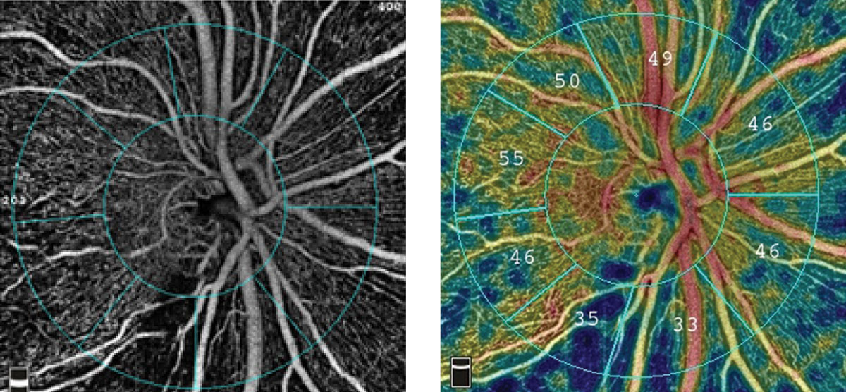 |
| In the present study, OCT-A-only progressors tended to have worse VF and thinner RNFL than OCT-only progressors. The researchers believed this implies that OCT-A may better reflect progressive changes as compared with OCT as glaucoma worsens. Photo: Visionix. Click image to enlarge. |
In this retrospective study, glaucoma eyes with ≥two-year and four-visits of OCT/OCT-A imaging were included. Peripapillary capillary density and retinal nerve fiber layer thickness (RNFL) were obtained from 4.5mm × 4.5mm optic nerve head (ONH) scans. Event-based OCT/OCT-A progression was defined as decreases in ONH measurements exceeding test-retest variability on ≥two consecutive visits. Visual field (VF) progression was defined as significant VF mean deviation worsening rates on ≥two consecutive visits.
Among 147 eyes (89 participants), OCT-A and OCT identified 33 (22%) and 25(17%) progressors, respectively. They showed slight agreement (kappa value = 0.06), with seven eyes (5%) categorized as progressors by both. When incorporating both instruments, the rate of progressors identified increased to 34%. Similar agreement was observed in diagnosis- and severity-stratified analyses.
Overall, more glaucoma eyes were found with progressive capillary density loss by OCT-A than with progressive RNFL loss by OCT, and there only was slight agreement between these two tests. Compared to progressors identified only by OCT, progressors identified only by OCT-A tended to have thinner baseline RNFL and worse baseline VF. VF progression was identified in 11 eyes (7%). OCT and VF showed fair agreement (kappa value = 0.26), with six eyes (4%) categorized as progressors by both. OCT-A and VF showed slight agreement (kappa value = 0.08), with four eyes (3%) categorized as progressors by both.
While all but one criterion found more eyes with progressive OCT-A change than with progressive OCT change, the difference in the numbers of OCT and OCT-A progressors varied across different cut-offs. The researchers believed “this indicates the capabilities and sensitivity of OCT and OCTA to detect progression by event-based method may still be influenced by the cut-offs used, and a gold standard should be established to facilitate its clinical application.”
“Future studies with longer follow-up are needed to further confirm the relationship of OCT-A changes with glaucomatous functional impairment,” the authors concluded in their paper.
Wu JH, Moghimi S, Nishida T, et al. Detection and agreement of event-based OCT and OCT-A analysis for glaucoma progression. Eye (Lond). November 11, 2023. [Epub ahead of print]. |
