 |
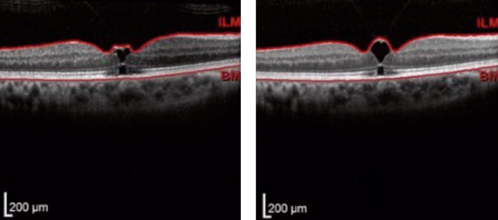
| Vitreomacular traction (shown here using conventional OCT methods) is one condition that may be better visualized and understood if a new OCT technique becomes refined and put into practice. Photo: Diana Shechtman, OD. Click image to enlarge. |
Clearly imaging the vitreous in vivo has been historically challenging for a number of safety and anatomical reasons, but now researchers have demonstrated a novel noninvasive method of enhanced vitreous imaging that they say provides insight into the vitreous architecture.
New York retina specialist Rick Spaide, MD, who essentially invented enhanced-depth imaging OCT some years ago by tinkering with his device, has now created a new software modification for the Heidelberg Spectralis to image the vitreous. So far, Dr. Spaide and colleagues from Switzerland have imaged just 10% of the posterior vitreous—admittedly a modest start, but one that does allow better visualization of the crucial vitreoretinal interface in vivo.
In this method, four A-scans are averaged four times before the Fourier transform step. The study authors noted in their paper that this boosting step provides sufficient detail to facilitate later frame averaging; however, it reduces scanning speed by a factor of four.
Despite this, they say scan speed remains tolerable with little opportunity for subject motion. “There are 0.0118 milliseconds between successive scans at the 85kHz sampling rate,” they noted in their paper.
To improve clarity, the authors defocused +2D from the retinal surface into the vitreous. They noted that the Spectralis requires sufficient overlap of strongly reflecting signals to align the successive OCT images. To ensure this, the retina and inner choroid must be in the image in the posterior pole.
The researchers obtained B-scans of 43 eyes (23 subjects) and averaged the scans 100 times. Upon analysis, they noted that the texture in the vitreous images suggested that the vitreous fibers in the macular region have specific orientations. Here are their findings:
Fibers were circumferential to the retina, immediately anterior to the premacular bursa.
Vitreous fibers had a less well-defined pattern internal to the zone of circumferential fibers.
Younger eyes had striations oriented in a roughly interior-to-superior direction.
Older eyes had striations in the same orientation but in alternating zones of vitreous synchysis and syneresis.
Additionally, they observed numerous cisterns (pockets of liquid vitreous) at various levels in the vitreous gel and noted that premacular bursa definition was lost with extensive vitreous condensation and synchysis. Images obtained in this study showed that the premacular bursa is surrounded by highly reflective vitreous gel. They suggest bursa formation may be the product of controlled vitreous condensation.
“The patterns of degeneration in the vitreous in our subjects appeared to follow stereotypic patterns, and these could potentially be modeled using numeric techniques,” the authors wrote.
They say this OCT boosting technique may aid in the advance of spectrometers for SD-OCT, SS-OCT and full-range imaging, since increased speed for data acquisition, storage and processing is needed. “Until that time, the present method used in this study provides significant improvements in imaging the 3D nature of the vitreous in living subjects,” the authors wrote.
Spaide RF, Valmaggia P, Maloca PM, et al. Imaging the vitreous with a novel boosted optical coherence tomography technique: vitreous degeneration and cisterns. Retina. March 17, 2022. [Epub ahead of print]. |
