A staggering two million new cases of keratitis are recorded each year. Although antibiotics are standard treatment, a more organism-specific therapy would be beneficial, but there is a lack of efficient and inexpensive diagnostic testing to detect specific keratitis pathogens in the office if one isn’t willing to wait for culture results to come back. In a new study, researchers investigated the diagnostic value of anterior segment OCT (AS-OCT) scans for Pseudomonas keratitis and found it’s a valuable tool in distinguishing the condition and has great potential in differentiating various pathogens causing keratitis.
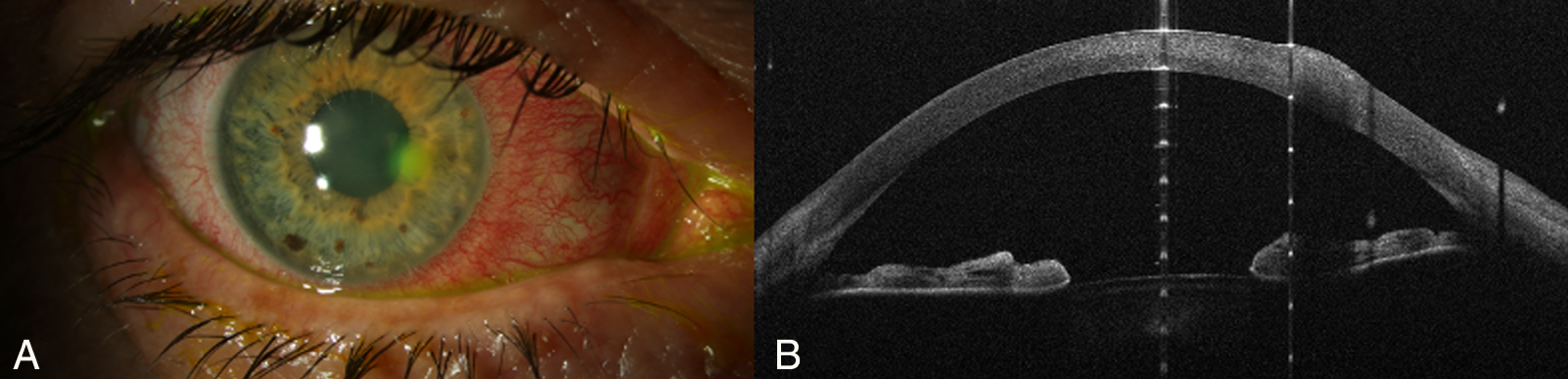 |
| A patient with contact lens–associated Pseudomonas keratitis and symptom onset within the last 24 hours. (A) Slit-lamp photo revealing a paracentral corneal infiltrate and conjunctival hyperemia. (B) AS-OCT demonstrates an increase in corneal thickness (tissue gain) at the lesion area. Photo: Khalil H, Bolz M, Waser K, et al. Trans Vis Sci Tech. Nov. 29, 2023. Click image to enlarge. |
A total of 19 patients with treatment-naïve keratitis underwent AS-OCT imaging— seven cases in the Pseudomonas group and 12 cases in the gram-positive group. Three different OCT devices were used to evaluate corneal thickness, infiltrate thickness, infiltrate diameter, tissue loss/gain, entropy and distance of the lesion from the corneal pupillary center. The relationship between the detected pathogen and the OCT patterns was analyzed.
A large infiltrate diameter and a gain of 30% of the corneal tissue appeared to be a predictive indicator for detecting Pseudomonas keratitis, while a gain of 15% was observed in cases with gram-positive bacteria.
“These results are in line with those of previous research, confirming that gram-negative bacteria display a larger surface area of infiltration,” the researchers wrote in a paper for Translational Vision Science & Technology. “A possible explanation for this may be the more severe inflammatory response of gram-negative infections due to certain bacterial virulence factors.”
The mean diameter from the Pseudomonas keratitis group was 2,067μm, much less from cases documented in the Steroids for Corneal Ulcer Trial (4,066μm and 3,061μm). Because patients were screened at an early phase of the infection, the authors suggested that the destructive enzymatic activity may not have yet fully manifested or caused extensive tissue damage.
“At this early stage, the infection might predominantly affect the corneal epithelium and anterior stroma, leading to localized changes that could be captured by AS-OCT imaging,” the authors explained in their article. “As a result, a tissue gain parameter could potentially reflect the initial inflammatory response and tissue edema, rather than the later-stage destructive changes. Additionally, the tissue gain parameter we describe is relative to the corneal thickness of the opposite corneal side. This accounts for the potential variability in corneal thickness among individuals and provides a standardized metric for assessing changes within the context of the individual’s own corneal thickness.”
The findings of this study could be used to develop new diagnostic strategies for Pseudomonas keratitis, the team suggests, as well as develop new biomarkers for the infection.
“Our study not only establishes the utility of AS-OCT in differentiating these bacterial etiologies based on quantitative measurements of infiltrate dimensions, corneal thickness and tissue loss/gain but also underscores its potential as a valuable prognostic tool,” the authors concluded. “By visualizing the structural changes within the cornea, AS-OCT can offer insights into the severity of tissue involvement and progression, which can guide prognostication and management decisions. In the future, a larger sample will be included and differences among several exotoxins will be evaluated.”
Khalil H, Bolz M, Waser K, et al. Diagnostic potential of anterior segment optical coherence tomography scans for Pseudomonas keratitis. Trans Vis Sci Tech. November 29, 2023. [Epub ahead of print.] |
