 |
|
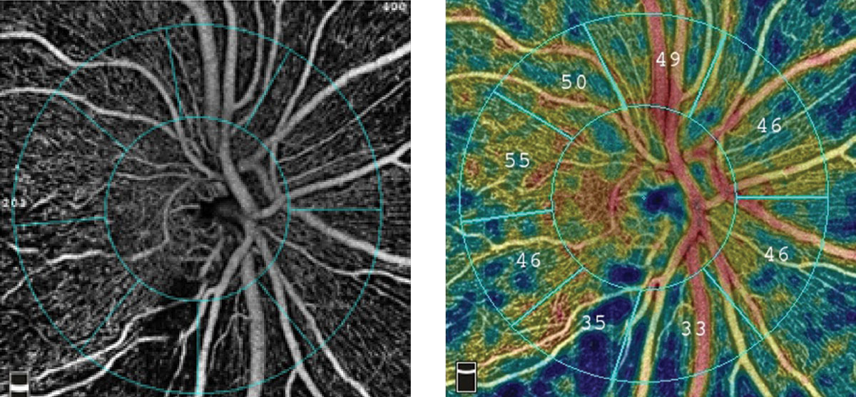
OCT-A measurements show that rates of macular VD loss were significantly faster in more advanced POAG eyes. Photo: Optovue. Click image to enlarge. |
Given the various stages of glaucoma, it’s important to understand the mechanism of disease progression. Previous studies using OCT angiography (OCT-A) in glaucomatous eyes have showed decreased vessel density (VD) in the macular area, so the purpose of this recent study was to characterize and compare the longitudinal changes of macular VD in primary open-angle glaucoma (POAG) eyes across different disease stages.
A total of 103 eyes (53 eyes in the mild stage, 50 eyes in the moderate-to-advanced stage) of 75 POAG patients were followed for more than one year with at least two OCT-A images. The rates of macular VD change were determined by linear regression and compared using generalized linear mixed models between groups. Mixed effect models were used to evaluate the demographic and ocular parameters associated with the VD loss rate.
The study demonstrated that significant macular superficial vasculature deterioration was detectable with OCT-A in POAG eyes. The rates of macular VD loss were significantly faster in more advanced stages of POAG. “Conversely, there was no significant visual field deterioration detected. The severity of visual field damage at baseline is significantly correlated with glaucomatous progression in terms of macular vasculature loss,” the authors explained in their paper.
The moderate-to-advanced group showed significantly lower whole-image macular VD at baseline compared with the mild group, which is consistent with prior studies that reported macular VD was significantly lower in eyes with advanced-stage POAG than in eyes with early-stage POAG.
When comparing VD across different sectors, eyes with moderate-to-advanced stages of glaucoma had lower VD values in the inferior hemifield, parafovea, inferior and temporal sectors compared with eyes with mild-stage glaucoma. This is again consistent with prior studies that evaluated the macular damage in glaucomatous eyes and reported the greatest differences between glaucomatous eyes and normal eyes in the inferotemporal sectors.
“In addition, we compared the longitudinal macular VD loss rates by sector between two groups. We found a statistically significant difference between the mild and moderate-to-advanced groups in the whole image, superior hemifield, parafovea, superior, nasal, inferior and temporal sectors,” the study authors explained. A possible explanation, they suggest, is that the inferior macular region was already damaged in the early stage of glaucoma, and there could be a lack of significant difference in longitudinal VD loss rates among different stages of glaucomatous eyes.
“In conclusion, the current study showed that OCT-A measurements could detect vascular deterioration in eyes with various stages of POAG,” the investigators explained. “The rates of macular VD loss were significantly faster in POAG eyes at a more advanced disease stage. The baseline disease stage is associated with the vascular progression of glaucoma in terms of macular VD loss. These findings suggest a possible advantage of applying OCT-A in monitoring glaucoma patients.”
Shang X, Wang X, Zhou K, et al. Faster macular vessel density loss in more advanced primary open angle glaucoma eyes. Ophthalmic Research. November 15, 2022. [Epub ahead of print]. |
