 |
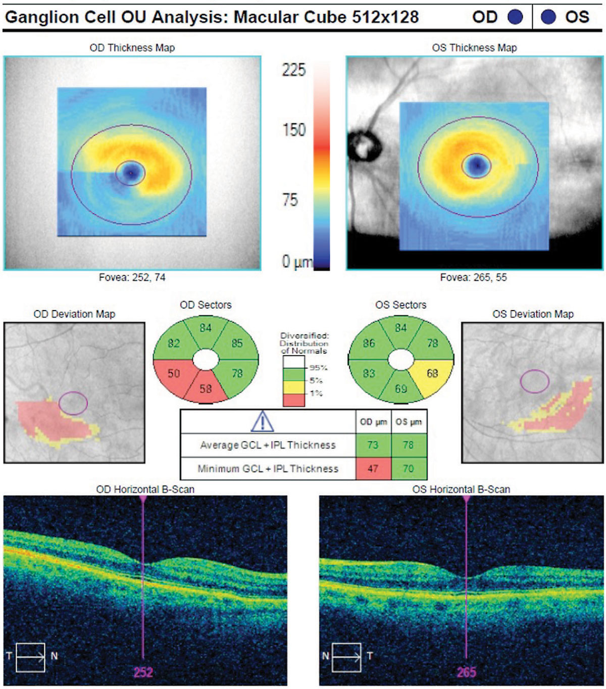
| While the study did not find a significant cross-sectional difference in GC-IPL thickness between patients with Parkinson’s and control patients, those with the condition had a significantly greater rate of GC-IPL thinning over time. (Image shows GC-IPL thinning in a glaucoma patient.) Photo: Danica Marrelli, OD. Click image to enlarge. |
There has been increasing effort to identify ophthalmic biomarkers to assist in both Parkinson’s disease screening and diagnosis as well as disease progression for which longitudinal data of imaging parameters is required. To clarify whether the rate of retinal and choroidal changes in patients with Parkinson’s is different from those attributed to normal aging, the Eye Multimodal Imaging in Neurodegenerative Disease (iMIND) Research Group based out of Duke University School of Medicine compared the rates of change with OCT and OCT-A. They observed higher rates of yearly loss, often multiple times higher, in vessel density, superficial capillary plexus perfusion density and ganglion cell-inner plexiform layer (GC-IPL) thickness in those with Parkinson’s disease compared with controls over a mean interval of 22.8 months. Two years is a relatively short span in the continuum of Parkinson’s.
The longitudinal study, published in Ophthalmology Science, compared 74 eyes of 40 patients with Parkinson’s disease and 149 eyes of 78 control individuals. Imaging parameters included central subfield thickness, GC-IPL thickness, peripapillary retinal nerve fiber layer (RNFL) thickness, choroidal vascularity index, superficial capillary plexus perfusion density, vessel density and foveal avascular zone area.
The findings of this study suggested that individuals with Parkinson’s may experience more rapid loss of microvasculature and faster rates of neurodegenerative decline in the retina when compared to controls. In addition, the velocity of decline may be greater in those with more severe disease. However, the research team noted in the paper that additional work with a larger sample size would be needed to confirm this observation.
Those with Parkinson’s had greater rate of yearly decrease in GC-IPL, greater yearly decline in plexus perfusion density in the 3x3mm ETDRS circle and ring, as well as the 6x6mm ETDRS circle and outer ring. Those same patients had greater rates of yearly decline in vessel density in the 3x3mm circle and ring, as well as the 6x6mm circle and outer ring.
“Over two years, there is a significantly greater velocity of decrease in superficial capillary plexus perfusion density, vessel density and GC-IPL thickness in participants with Parkinson’s disease compared with controls,” the researchers wrote in their paper.
“Incorporation of retinal and choroidal parameters as a supporting target in clinical trials investigating disease-modifying approaches in Parkinson’s, while promising, requires further validation of these biomarkers,” they added. “Additional investigation will determine whether the rate of change slopes of these parameters have a role in biological stratification of disease progression.”
Kundu A, Ma JP, Robbins CB, et al. Longitudinal analysis of retinal microvascular and choroidal imaging parameters in Parkinson disease compared to controls. Ophthalmol Sci. September 4, 2023. [Epub ahead of print]. |
