 |
|
Researchers have yet to learn whether retinal microvasculature changes are a cause or a consequence of amblyopia but have at least documented an association for now. Photo: Julie Rodman, OD. Click image to enlarge. |
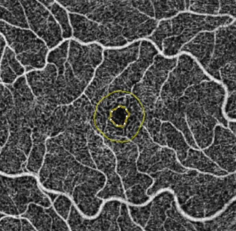
Evaluating the microvascular system is an intriguing new avenue to potentially diagnose amblyopia in patients. OCT angiography (OCT-A) is a noninvasive imaging modality that can provide quantitative blood density or flow data within the retina and choroid without the need for injection of fluorescein dye. A recent study’s data suggests that whole foveal, parafoveal and radial peripapillary capillary whole vessel density (VD) were reduced in patients with amblyopia. Moreover, the results revealed that the deep foveal avascular zone (FAZ) was larger in amblyopic patients. Consequently, the researchers concluded that OCT-A may have the potential to diagnose and monitor patients with amblyopia.
The team analyzed 1,832 eyes (353 in the amblyopia group and 1,479 in the control group). They evaluated several outcomes relevant to diagnosing amblyopia, including macular whole en face VD, parafoveal VD, radial peripapillary capillary whole en face VD and FAZ.
Compared with controls, the foveal whole en face VD of the superficial and deep capillary plexus of patients as measured by 3x3mm scans was significantly lower in amblyopia eyes (-1.37 vs. -1.70). Similarly, in the 6x6mm scans, the foveal whole en face VD of the superficial and deep capillary plexus was remarkably lower in amblyopia eyes than controls (-2.24 vs. -5.08). The parafoveal VD of the superficial capillary plexus in 3x3mm scans (-1.96) was also lower in amblyopic patients than in controls. In 6x6mm scans, amblyopia eyes showed a significant decrease, as well as a trending decrease in the parafoveal VD of the superficial and deep capillary plexus (-3.85 vs. -3.03).
For whole radial peripapillary capillary, the VD was significantly reduced in amblyopic patients compared with controls (-0.83). In addition, the deep FAZ was larger in amblyopic eyes than controls (0.55).
Whether a decrease in retinal microvascular density is the cause or consequence of amblyopia remains unknown. The researchers noted that further longitudinal studies should explore the in-depth pathophysiology of the changes in microvascular density in eyes with amblyopia.
“Our work suggests that OCT-A represents a promising device for objectively diagnosing and monitoring amblyopia,” they concluded in their paper.
Yang CC, Ji KB, Yu YF. Analysis of retinal microvasculature features in amblyopic eyes: a meta-analysis. Ophthalmic Res. August 23, 2022. [Epub ahead of print]. |
