 |
Q:
I have a patient who needs a lamellar graft for endothelial dysfunction but has previously had a vitrectomy for a retinal detachment. Can this patient have a Descemet’s membrane endothelial keratoplasty (DMEK) procedure to restore corneal integrity or should a different form of lamellar surgery be used?
Descemet’s stripping automated endothelial keratoplasty (DSAEK) and DMEK have improved nearly every aspect of the immunologic and optical impact of penetrating keratoplasty for patients with endothelial disease. “Despite these improvements, both present unique challenges, particularly intraoperatively and in the early postoperative period. Prior ophthalmic surgery in some cases can add dramatic complexity to the case,” says Aaron Bronner, OD, of Pacific Cataract and Laser Institute in Boise, ID.
Of particular concern are previous vitrectomy, filtering tubes or trabeculectomies, large surgical iridectomies or any procedure or trauma resulting in a significant compromise to the zonulo-capsular complex and aphakia.
Background
Understanding how these surgeries potentially complicate DSAEK or DMEK requires a bit of understanding about the surgical process of each. Dr. Bronner explains that in both transplant types, the central host Descemet’s membrane and endothelium is scored and carefully removed. The graft is then introduced into the anterior chamber. In DSAEK, the graft generally unfolds easily with injection of a balanced salt solution.
With DMEK, the graft is scrolled, and getting it to un-scroll is a bit of a trick, as the surgeon can’t simply grab it and unroll it—excessive direct touch will often cause the graft endothelium to fail. Instead, the surgeon relies on percussive waves and balanced salt solution flow through the anterior chamber to get the graft to un-scroll. The surgeon facilitates this by somewhat shallowing the anterior chamber, as surface tension interactions between both the host cornea and iris with the graft assist with its unrolling.
 |
|
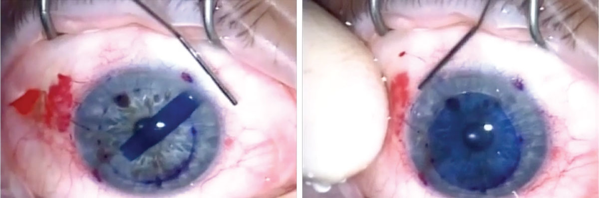
In this photo sequence, cornea surgeon Jim Guzek, MD, is unrolling a DMEK graft. The graft is stained with Trypan Blue to aid visualization of the very thin tissue. Through circulating BSS and creating percussive waves through the cornea, with the aid of surface tension interactions of the graft, host cornea and iris, he is able to get the tightly scrolled transplant to unroll without touching it. Click image to enlarge. |
Once the graft is in position, an air or air/gas mix is injected into the anterior chamber and the eye is pressurized (and chamber deepened) for a period in surgery and the patient is observed.
After some time, the eye is depressurized through a bubble (of variable size and make-up, depending on the surgery) is left in place, Dr. Bronner describes. The patient is sent home with positioning restrictions to remain supine for much of the following days. This allows the bubble to press the graft into place and, if the transplanted endothelium is viable, will allow the graft to remain in position as the bubble re-absorbs.
Complications
Vitrectomy can create two particular issues for DMEK. First, the anterior chamber and vitreal cavity are no longer separate pressure systems; second, the ability to shallow the anterior chamber to aid in the unrolling of the graft is inhibited. This problem is specific to DMEK and does not apply to DSAEK. Secondly, if the patient also has a compromise to the capsule or zonules, the air/gas bubble can escape posteriorly and may remain stuck behind the intraocular lens.
Without a predictable air/gas bubble tamponade, the graft is likely to detach. This can be a problem for both DSAEK and DMEK. “Not all vitrectomized eyes have compromised capsular/zonular integrity, so this issue varies by individual,” Dr. Bronner notes. “Large superior surgical iridectomies and scleral fixated IOLs may also complicate these transplants due to posterior migration of the air bubble,” he adds.
Filtering surgeries create issues with the ability to pressurize the eye after placement of the graft. If the eye can’t be pressurized appropriately, it is much more difficult to get the graft to adhere prior to sending the patient home. Further, eyes with glaucoma severe enough to justify filtering surgery may not be good candidates for DSAEK or DMEK because of attempts that will be made to pressurize the eye and the impact that may have on their remaining retinal nerve fiber layer.
According to Dr. Bronner, aphakia complicates DSAEK and DMEK. Though you can imagine getting a bubble to stay in the anterior chamber may be difficult in a patient with aphakia, usually these patients have already had vitrectomies, so the entire eye can often be filled with gas/air. Instead, the primary issue is not being able to easily shallow the chamber (as with simple vitrectomy) and the potential for the transplant dropping to the retina intraoperatively.
Descemet’s stripping only may be used to avoid these issues, however, the surgery is niche. Descemet’s stripping only is limited to patients who have endothelial disease from Fuchs’ dystrophy (no other source of endothelial decompensation can be treated with this surgery), and it tends to work best in those who primarily have central involvement, so isn’t an option in many cases of endothelial decompensation.
“None of these postoperative states create problems that are entirely insurmountable, but cases like this may exceed the comfort or ability of community cornea specialists who may perform only a couple transplants a month,” concludes Dr. Bronner. “Centers that specialize in transplants specifically may be better suited to tackle cases that are anticipated to be challenging.”
Dr. Shovlin, a senior optometrist at Northeastern Eye Institute in Scranton, PA, is a fellow and past president of the American Academy of Optometry and a clinical editor of Review of Optometry and Review of Cornea & Contact Lenses. He consults for Kala, Aerie, AbbVie, Novartis, Hubble and Bausch + Lomb and is on the medical advisory panel for Lentechs.