 |
|
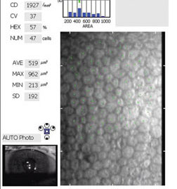
Study shows there is a significant relationship between cell size and duration of diabetic with age variable control. Photo: Francis W. Price Jr., MD. Click image to enlarge. |
Clinicians most often think of diabetic retinopathy (DR) risk in their diabetes patients but the condition can also affect the anterior segment, including the corneal endothelium. Due to limited studies on the association, authors of a recent study investigated the potential link in type 2 diabetes patients.
A total of 134 eyes of 134 diabetic patients were enrolled and split into five subgroups: diabetic patients without retinal involvement, diabetic patients with mild and moderate nonproliferative DR (NPDR), diabetic patients with severe NPDR, diabetic patients with mild proliferative DR (PDR) and diabetic patients with high-risk PDR. Corneal endothelium parameters including endothelial cell density, average cell size, coefficient of variation in cell size and hexagonality were evaluated by non-contact specular microscopy.
The study demonstrated that diabetes has negative effects on cell size as one of the important corneal endothelium parameters. There is a significant relationship between the cell size and the duration of disease with age variable control that is a “predictable and acceptable finding regarding increasing the adverse effect of diabetes in all organs with increasing the duration of diabetes,” the authors explained. This may be due to differences in ethnicity and duration of disease in the enrolled population, they added.
A possible explanation for corneal endothelial changes in diabetes patients is multifactorial, the authors explained, including impairment of apical junctions on the endothelial cells, impairment of physical barriers of corneal cells and altered permeability of corneal cells due to reduced Na+/K+ ATPase activity pump in the endothelial cells. “A diabetic cornea, especially with high glucose, can lead cellular swelling due to increased sorbitol inside the cells due to increased activity of aldose reductase,” the investigators wrote.
Since there was no endothelial cell proliferation with age, the number of corneal endothelial cells will decrease, the authors noted. To make up for this shortcoming, existing cell size will increase to compensate for the lost cells.
“In each ocular surgery, the cell size number should be in the normal range to ensure that decompensation does not occur after the ocular surgery,” the authors explained. “In the current study, it was shown that there were no significant differences in cell size number in NPDR and PDR groups.”
Because of the relationship between cell size and the duration of the disease with age variable control, the authors suggest long-lasting DM may warrant a further corneal evaluation before intraocular surgery.
Mortazavi SAA, Akhlaghi M, Dehghani A, et al. Diabetic retinopathy and corneal endothelial parameters: an analytical cross-sectional study. BMC Ophthalmol. November 9, 2022. [Epub ahead of print]. |
