 |
| Retinal vascular injury may predict cognitive impairment, as this study and previous ones suggest. Photo: Carolyn Majcher, OD. Click image to enlarge. |
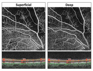
The retina has been proposed as a potential biomarker of cognitive impairment for some time now. In a new study performed in China, the relationship between the structures of the retinal capillary plexus (RCP) and ganglion cell complex (GCC) with mild cognitive impairment (MCI) and dementia was explored.
OCT angiography was performed to obtain RCP vessel density and GCC thickness. Cognitive status was assessed by professional neuropsychologists using the Mini-Mental State Examination and Montreal Cognitive Assessment. Subjects were divided into three groups: normal, MCI and dementia.
Among the 2,678 participants, MCI was found in 7.4% (n=197) and dementia in 3% (n=80). Only decreased deep RCP vessel density was found to be associated with MCI, while dementia was associated with decreased superficial and deep RCP, as well as a thin GCC. Looking deeper into the association of RCP and MCI, the researchers noted alterations according to age, alcohol consumption and education.
The observed decreased deep plexus vessel density was consistent with many previous studies. Many found that Alzheimer’s patients and mouse models display amyloid beta plaque accumulation that is detectable earlier in retinal vessels than brain structures. Another study showed that changes in retinal oxygen saturation in MCI patients preceded Alzheimer’s disease.
This study further explores the association by suggesting deep retinal, vascular pathological changes occur in early stages of cognitive impairment. The researchers, in their paper for Eye, explain one reason for this might be that the deep capillary plexus is more sensitive to hypoxia. They noted that “at the MCI stage, deep capillary plexus damages might occur due to the cardiovascular risk factors of cognitive impairment, but the GCC could still receive sufficient blood supply from the superior capillary plexus because of the location of the superior plexus in the retinal nerve fiber layers and ganglion cell layers.”
Similarly, the structures associated with dementia here were similar to prior study findings. A thinner ganglion cell and inner plexiform layer is associated with less white matter microstructure integrity and less gray matter volume in the cerebellum and visual cortex. As such, the researchers believe this may indicate a thinner GCC reflects central nervous system degeneration in advanced cognitively impaired patients.
In dementia, compared with earlier stages of cognitive impairment, both superior and deep capillary plexus damage involvement negatively affected the GCC, which may in turn be associated with phenotypic brain changes.
Comparing demographics, an association between MCI and the superior capillary plexus was observed in patients younger than 60. Alcohol consumption strengthened the association between the MCI and the deep capillary plexus, thus showing RCP loss is more serious in MCI patients who consumed alcohol. As such, the authors believe “we should pay special attention to RCP loss in young individuals who consume alcohol due to its interactive effects on the association with MCI,” they wrote. Finally, higher education was more strongly associated with deep capillary plexus and MCI than lower education.
Clinically, the authors suggest, the “deep capillary plexus provides possibilities for exploring sensitive retinal markers to detect MCI earlier, as well as a decreased superior capillary plexus and deep capillary plexus and thin GCC for dementia.”
Li C, Zhu X, Yang K, et al. Relationship of retinal capillary plexus and ganglion cell complex with mild cognitive impairment and dementia. Eye. June 2, 2023. [Epub ahead of print]. |
