 |
A patient who schedules an appointment for a red eye could walk through the door with any number of ocular issues. This month’s feature articles hit the highlights: allergy, infection, dry eye, scleritis and episcleritis, to name only a few. But some patients still call their primary care provider for a red eye, especially if the patient is a child. Many parents aren’t aware of where to take their children when they have ‘pink eye.’
We must continue to educate patients—starting with each parent or patient who enters our offices—on our services beyond the basic refraction that once defined us. We have to directly or indirectly convey to our patients that we manage anything related to the eyes, including “red eyes” and children. In addition to managing garden-variety conjunctivitis most effectively, we can also ensure that it is not an iritis, preceptal cellulitis, episcleritis, keratitis or some other potential differential diagnosis that could be more painful and problematic.
Optometrists also know to look for systemic findings in patients who present with conjunctivitis. The most common causes of bacterial conjunctivitis in children, for example, include Haemophilus influenza and Streptococcus pneumonia. Unlike adult conjunctivitis, which is most commonly caused by Staphylococcus, the pathogens in childhood conjunctivitis cause a significant number of systemic issues, including otitis, preceptal cellulitis and, in rare cases, encephalitis.
The first step when examining a child with conjunctivitis is checking their temperature and questioning the parents regarding systemic findings. A fever, ear infection, malaise or upper respiratory infection warrants systemic treatment, and only then would a referral to the pediatrician be best. The presence of a reddish sheen around the eyes indicates preceptal cellulitis and requires oral antibiotics in addition to topical treatment—something optometrists can handle.
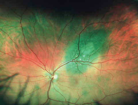 |
| Although this patient had no complaints, his optometrists found a large retinal lesion during the dilated exam, later diagnosed as malignant choroidal melanoma. |
Beyond the Ocular Surface
Now is a critical time for optometry to educate the masses. It is always in our interest to educate about the technology we use and the conditions we can treat, including everything from a common conjunctivitis to the most critical and rare findings. It may be ‘pink eye’ or another basic issue that brings them in; once they’re in the chair, however, we can truly play a life-changing role.
Take a recent 50-year-old patient. He presented to a local optometrist simply because of symptoms related to presbyopia. He measured 20/20+ OD and OS. Because it was his first exam with an optometrist, he was dilated and received a comprehensive exam. The OD captured the image provided here, and immediately referred him to our retina specialty practice for further evaluation. Fundus exam revealed a large lesion, OCT imaging confirmed the presence of serous fluid, autofluorescence confirmed lipofuscin over the lesion and ultrasound confirmed a diameter of more than 3mm. All of this led to a confirmed diagnosis of a malignant choroidal melanoma.
Though he was referred to our specialty practice, every one of these tests could have been conducted by the primary eye care provider. OCTs are commonplace in optometry, as are ultra-widefield imagers and dilated fundus exams. Even B-scan ultrasounds are now inexpensive and have great image resolution.
This is just one more example of why it is so important to educate our patients about our knowledge, training and capabilities. Without optometric intervention, this melanoma would surely have resulted in devastating consequences.
