| |
|
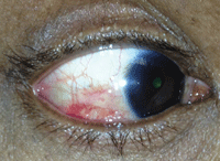
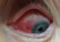
Case 1. Sectoral nodular scleritis.
|
Scleritis can be classified as either anterior or posterior, where anterior scleritis can be further categorized into four different types:
• Diffuse scleritis is widespread inflammation of the sclera, and the most common type.1
• Nodular scleritis is characterized by a localized area of inflammation where a distinct nodule can be seen.
• Necrotizing scleritis with inflammation is frequently associated with collagen vascular disorders causing destruction of the sclera.
• Necrotizing scleritis without inflammation, also known as scleromalacia perforans, is a very rare form of scleritis presenting with no symptoms. It can be seen in patients with rheumatoid arthritis, granulomatosis with polyangiitis (formerly known as Wegener’s granulomatosis) and relapsing polychondritis.2
|
|
|
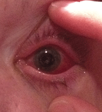
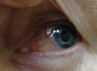
Case 2. Scleral inflammation in a patient who had recent angle-closure glaucoma.
|
Posterior scleritis can also be nodular or diffuse and necrotizing, and involves the sclera posterior to the insertion of the rectus muscles.2
In this article, we present a few challenging cases of scleritis to highlight various treatment approaches and learning points.
Case 1. Red Eye, Cough and Weight Loss
• History. A 66-year-old Indian male presented with bilateral red and mildly painful eyes. He said his eyes had been getting red on and off, but this episode was much worse.
• Diagnostic data. Slit-lamp exam showed bilateral sectoral nodular scleritis. Although it was mostly superficial, there was some deeper episcleral inflammation noted. His vision was correctable to 20/25- OD and OS, and his intraocular pressures were 18mm Hg OD and 19mm Hg OS.
Upon questioning, he reported chronic respiratory issues with a persistent cough for the past four to six months. He had been diagnosed with recurring bronchitis and was treated with several courses of antibiotics as well as oral steroids. He had also lost an unexplained 20 lbs. during the past four months and was reporting generalized muscular pain.
He was already taking a course of prednisone for his respiratory issues, so started him on Durezol (difluprednate 0.05%,
Alcon) QID. Given his bilateral scleral inflammation along with his systemic issues, we ordered additional lab testing, which included: RF, ANA, HLAB27, Lyme, PPD, FTA-ABS, ACE, C-ANCA, P-ANCA, ESR, CRP. (See “Common Laboratory Tests for Patients with Scleritis,” right.)
|
Common Laboratory Tests for Patients with Scleritis
| |
|
Laboratory Test
|
Systemic Condition
|
|
ACE (angiotensin-converting enzyme)
Chest X-ray |
Sarcoidosis
|
|
ANA (antinuclear antibody)
|
Lupus
|
|
c-ANCA (cytoplasmic antineutrophil cytoplasmic antibody)
|
Wegener's granulomatosis
|
|
p-ANCA (perinuclear antineutrophil cytoplasmic antiybody)
|
Vasculitis, polyarteritis nodosa
|
|
FTA-ABS (fluorescent treponemal antibody absorption)
RPR/VDRL (rapid plasma reagin/venereal disease reference laboratory) |
Syphilis
|
|
ELISA (enzyme-linked immunosorbent assay)
Western blot |
Lyme disease
|
|
RF (Rheumatoid factor)
|
Rheumatoid arthritis
|
|
CRP (C-reactive protein)
ESR (erythrocyte sedimentation rate) |
Nonspecific systemic inflammation
|
When he returned one week later, his eyes were white and quiet. We reviewed his lab results, which showed significantly elevated ANCA and CRP levels. We contacted his pulmonologist due to a high concern for granulomatosis with polyangiitis (GPA, aka Wegener’s granulomatosis); imaging studies and a lung tissue biopsy later confirmed the diagnosis. Our patient was sent to a GPA specialist for further evaluation and treatment.
• Discussion. Half of all scleritis cases have an underlying systemic cause.2 So, a thorough history and review of systems are very important to help identify any underlying etiologies and guide laboratory studies or further medical evaluation.
GPA is often overlooked due to its rare incidence—just three cases per 100,000 people.3 Approximately 10% of patients with GPA develop scleritis, so ANCA testing should be included in the workup of these patients.4
In addition to GPA, rheumatoid arthritis, relapsing polychondritis, polyarteritis nodosa, systemic lupus erythematosus, sarcoidosis, anklyosing spondylitis herpes zoster, syphilis and tuberculosis are associated with scleritis and should be tested as causative factors.2
Our patient returned for follow-up six months later. He was tapering prednisone and had started methotrexate for the GPA. The overall improvement in his health was remarkable. He had gained back 20 lbs., and was feeling significantly better with no more breathing or coughing issues. His scleral inflammation has been quiet since.
Case 2. Angle Closure or Inflamed Sclera?
• History. A 47-year-old white female presented for a second opinion with a very painful, red left eye with decreased vision. She had been diagnosed with primary angle-closure glaucoma and had undergone a YAG peripheral iridotomy a few days prior to this visit. However, after the procedure, she was still in a moderate amount of pain, which was worsening.
|

|
|
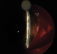
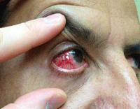
Case 2. A very shallow anterior chamber due to posterior scleritis. Inflammation has caused the ciliary body to rotate, creating anterior displacement of the lens iris diaphragm.
|
After cyclopegia, the ciliary body has re-rotated back to its original position and the angles have deepened. Also, anti-inflammatories have reduced ciliary body edema.
|
She was taking Pred Forte (prednisolone 1%, Allergan) QID, Azopt (brinzolamide 1%, Alcon) BID and Combigan (brimonidine 0.2%/timolol 0.5%, Allergan) BID.
• Diagnostic data. Her visual acuity was 20/20 with plano correction in the right eye and 20/100 in the left eye, which improved to 20/30 with a -2.00D correction. Her corneas were clear. Left anterior segment showed moderate conjunctival/scleral erythema, a 2+ anterior chamber cell and a very shallow anterior chamber with a patent peripheral iridotomy.
Gonioscopy in the left eye showed no visible angle structures. Her right anterior segment was normal with a deep anterior chamber and gonioscopy open to her ciliary body. Her intraocular pressure measured 18mm Hg OD and 42mm Hg OS. Her retinal exam showed an annular choroidal detachment.
• Discussion. Given the open angle in her right eye, absence of risk factors for angle closure (such as hyperopia or a denser cataract), poor response to YAG PI, myopic shift and choroidal detachment, we determined that her primary issue was posterior scleral inflammation causing a ciliochoroidal effusion syndrome and not pupillary block angle closure.
Posterior scleritis with ciliary body inflammation can initiate ciliary body rotation, which causes an anterior displacement of the lens iris diaphragm resulting in secondary angle closure.5 Additionally, ciliary body edema relaxes the lens zonules, which causes thickening of the crystalline lens and a myopic shift, as in this patient.6
Ciliochoroidal effusion syndrome, although rare, can often be mistaken for pupillary block angle closure. It is incredibly important to be able to distinguish the two conditions because the treatments are vastly different. Pupillary block glaucoma is typically seen in older individuals who have denser cataracts.7 Hyperopes are at a greater risk due their shorter axial length and shallower anterior chamber depth.7 Pilocarpine, steroid drops and IOP-lowering agents, along with a peripheral iridotomy, help resolve pupillary block glaucoma. In contrast, ciliochoroidal effusion syndrome can be reversed by cycloplegia along with anti-inflammatories and IOP-lowering agents. The anti-inflammatories reduce ciliary body edema and cycloplegia induces posterior rotation of the ciliary body, deepening the anterior chamber.6,8
We cyclopleged the patient in office, discontinued Pred Forte and started her on Durezol QID, homatropine 5% BID, oral prednisone 60mg, and continued Combigan and Azopt.
At one-day follow-up, there was a significant improvement of inflammation, and examination revealed a deepening of the anterior chamber. The patient’s vision improved to 20/30+ OS and the IOP reduced to 16mm Hg. We continued to manage her on anti-inflammatories and slowly tapered them as well as her IOP meds.
Laboratory testing showed an elevated antinuclear antibody, which prompted us to send the patient for rheumatologic evaluation. The rheumatologist diagnosed her with lupus.
At her last follow-up, her inflammation had resolved, IOP was stable, anterior chamber was deep and the choroidal detachment had resolved.
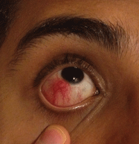
Case 3. Cancer Med is a Confounder
• History. A 42-year-old white male presented with an acute, painful, red right eye that had started the day before. He had a systemic history of melanoma with brain metastasis for which the primary lesion had not been identified. Previous ocular examinations were negative for choroidal or iris malignancies. The patient recently underwent removal of a metastatic brain lesion and was started on Zelboraf (vemurafenib, Genentech) a protein kinase inhibitor used to treat late-stage melanoma.9
| |
|
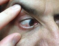
Case 3. A similar patient who presented with nodular, non-necrotizing scleritis. Treatment involved Durezol QID and a Medrol Dosepak PO. At one-week follow up, the scleral inflammation had resolved.
|
• Diagnostic data. Entering visual acuity was 20/25 OD and 20/20 OS. IOPs were within normal limits at 15mm Hg OD and 16mm Hg OS. There was a moderate nodular, non-necrotizing scleritis in the right eye. After instilling phenylephrine drops, moderate scleral inflammation was still noted temporally and inferiorly. There was a trace anterior chamber cell in that eye as well. The left eye was unremarkable.
We started the patient on Durezol QID and a Medrol Dosepak (methylprednisolone 4mg, Pfizer) PO. At one-week follow up, the scleral inflammation had resolved. We instructed the patient to taper the Durezol over the next two weeks.
• Discussion. Be aware of possible ocular effects from systemic medications. A thorough review of current medications is just as important as a review of symptoms. Although a rare side effect, ocular inflammation associated with Zelboraf has been documented and should be considered because it is being used more commonly for treatment of late-stage melanoma.9,10 In one report, Zelboraf was associated with a case of severe sclerouveitis.9 The drug has also been reported to cause uveitis in up to 4.5% of patients.10
|
Scleritis vs. Episcleritis
|
|
|
Scleritis can be differentiated from episcleritis both by history and clinical examination.
• Episcleritis typically presents with little to no pain and is most often idiopathic. It is generally a self-limiting inflammatory process affecting only the conjunctival and superficial episcleral vascular plexuses.2 • Scleritis patients, by contrast, present with moderate to severe pain and the inflammation extends into the deep episcleral plexus. Phenylephrine can be instilled in the eye to determine the depth of the inflammation. The conjunctival and superficial vessels will blanch or constrict from phenylephrine administration, but the deeper episcleral vessels will not. Treatment of both episcleritis and scleritis is aimed toward controlling inflammation. Episcleritis can be treated with topical steroids or oral NSAIDs along with lubricants, while scleritis is managed with both topical and oral anti-inflammatories. |
|
|
|
|
To help differentiate episcleritis from scleritis, use phenylephrine to blanch the superficial vessels—the deeper, episcleral vessels won't blanch.
|
|
In addition to Zelboraf, bisphosphonates—commonly used for treatment of osteoporosis—can also be associated with scleritis.11
In this patient’s case, comanagement with oncology was critical. We kept in close contact with the oncologist and, because of the patient’s good response to treatment with no additional recurrences, he continues to take Zelboraf and is monitored closely.
We did order a rheumatologic evaluation as well, just to rule out the possibility of underlying systemic etiologies, which was negative.
Case 4. Hit This Patient Hard and Fast (with Steroids)
• History. A 42-year-old Hispanic male was referred to our office for a painful, red right eye for the past three weeks that was unresponsive to treatment and was developing elevated intraocular pressure. The patient had been treated with Lotemax (loteprednol 0.5%, Bausch + Lomb) QID during this time but had no improvement.
• Diagnostic data. Entering visual acuity was 20/30 OD and 20/20 OS. Intraocular pressure was 25mm Hg OD and 15mm Hg OS. Slit-lamp examination showed a moderate nodular anterior scleritis in the right eye. Retinal exam was unremarkable in both eyes.
We switched the treatment to Durezol QID and a Medrol Dosepak. We also prescribed Combigan and Azopt to treat the steroid-induced ocular hypertension.
One week later, the patient returned for follow-up with a significant reduction in scleral inflammation and improvement of symptoms. Intraocular pressure was reduced to 20mm Hg in the affected eye. The patient finished taking the methylprednisolone and was switched from Durezol to Pred Forte for taper over the next 10 days.
|
|
|
This patient presented with a moderate nodular anterior scleritis, which was unresponsive to Lotemax, as well as elevated intraocular pressure.
|
After one week of treatment with Durezol QID and a Medrol Dosepak, as well as aqueous suppressants for the IOP, the inflammation had significantly improved.
|
At the following visit one week later, his scleral inflammation had completely resolved and his intraocular pressure was stable at 18mm Hg in the affected eye. We discontinued all drops at that time.
• Discussion. Although loteprednol can be used to treat milder cases of episcleritis (especially if there is a concern with a patient’s intraocular pressure), more potent steroid drops should be used for treating scleritis. (See “Scleritis vs. Episcleritis,” page 29.)
Clearly, one of the advantages of loteprednol over prednisolone, dexamethasone and difluprednate is the lower incidence of steroid-induced ocular hypertension. As prednisolone and difluprednate are ketone-based steroids, the molecules will linger in the eye longer.12 Loteprednol is an ester-based steroid, so any unbound molecules become metabolized by the natural esterases within the eye.12 As a result, loteprednol has a lower incidence of steroid-induced glaucoma; however, the risk still exists, as seen in our patient.
When treating scleritis, we typically take the “hit ’em hard, hit ’em fast” approach. This generally minimizes the duration of steroid therapy. We find it better to start off with a more potent corticosteroid drop and switch to a milder one, if needed, after a good amount of the inflammation has resolved.
|
Scleritis Responds to Oral Anti-Inflammatories
|
|
In addition to topical steroid drops, oral NSAIDs or oral steroids are
indicated for treating scleritis. Ibuprofen and indomethacin are often
used initially for treating anterior diffuse and nodular scleritis.
For mild scleritis cases, consider also prescribing a 4mg Medrol Dosepak (methylprednisolone, Pfizer). The self-tapering schedule helps with ease of use and patient compliance. For more moderate to severe cases, start with 60mg to 80mg of prednisone, and taper accordingly. |
Steroid drops administered for four to six weeks can cause an IOP rise of 6mm to 15mm Hg in 30% of patients and more than 16mm Hg in 5% of patients.13,14 Our goal of treating aggressively from the start is to have most patients off steroid therapy before this time frame. Even with moderate scleritis in a patient who is a known steroid responder, we will still treat aggressively and pre-treat with IOP drops in anticipation of the steroid response.
The diagnosis, treatment and management of scleritis can vary based on presenting signs, symptoms and associated factors. Anti-inflammatory agents are key in treatment of this condition, and should be used quickly and appropriately to maximize patient response. Also, be sure to consider all aspects of the patient’s medical history and review of systems in order to initiate appropriate therapy. Good communication between optometrists, internists and other medical specialists help to facilitate prompt diagnosis and adequate care of any underlying disease.
Dr. Trottini is in practice at Outlook Eyecare, in Monroe Township, NJ.
Dr. Tolud is in practice at South Jersey Eye Physicians, in Columbus, NJ.
1. Sims J. Scleritis: presentations, disease associations and management. Postgrad Med J. 2012 Dec;88(1046):713-8.
2. Okhravi N, Odufuwa B, McCluskey P, Lightman S. Scleritis. Surv Ophthalmol. 2005 Jul-Aug;50(4):351-63.
3. Tarabishy AB, Schulte M, Papaliodis GN, Hoffman GS. Wegener’s granulomatosis: clinical manifestations, differential diagnosis, and management of ocular and systemic disease. Surv Ophthalmol. 2010 Sep-Oct;55(5):429-44.
4. Hoffman GS, Kerr GS, Leavitt RY, et al. Wegener’s granulomatosis: an analysis of 158 patients. Ann Intern Med. 1992 Mar 15;116(6):488-98.
5. Ugurbas SH, Alpay A, Ugurbas SC. Posterior scleritis presenting with angle closure glaucoma. Ocul Immunol Inflamm. 2012 Jun;20(3):218-20.
6. Litwak AB. Posterior scleritis with secondary ciliochoroidal effusion. J Am Optom Assoc. 1989 Apr;60(4):300-6.
7. Lee JW, Lai JS, Lam RF, et al. Retrospective analysis of the risk factors for developing phacomorphic glaucoma. Indian J Ophthalmol. 2011 Nov-Dec;59(6):471-4.
8. Ikeda N, Ikeda T, Nomura C, Mimura O. Ciliochoroidal effusion syndrome associated with posterior scleritis. Jpn J Ophthalmol. 2007 Jan-Feb;51(1):49-52.
9. Wolf SE, Meenken C, Moll AC, et al. Severe pan-uveitis in a patient treated with vemurafenib for metastatic melanoma. BMC Cancer. 2013 Dec 1;13:561.
10. Sandhu S, Ling C, Lim L, et al. Vemurafenib (B-RAF inhibitor) associated uveitis in patients with metastatic cutaneous melanoma. Clin Exp Ophth. 2012;40(S1):118.
11. Leung S, Ashar BH, Miller RG. Bisphosphonate-associated scleritis: a case report and review. South Med J. 2005 Jul;98(7):733-5.
12. Comstock TL, Decory HH. Advances in corticosteroid therapy for ocular inflammation: loteprednol etabonate. Int J Inflam. 2012;2012:789623.
13. Armaly MF. Statistical attributes of the steroid hypertensive response in the clinically normal eye. I. The demonstration of three levels of response. Invest Ophthalmol Vis Sci. 1965 Apr;4:187-97.
