 |
The results of this study could be used as the foundation for machine learning training to automate OCT biomarker identification. Click image to enlarge. |
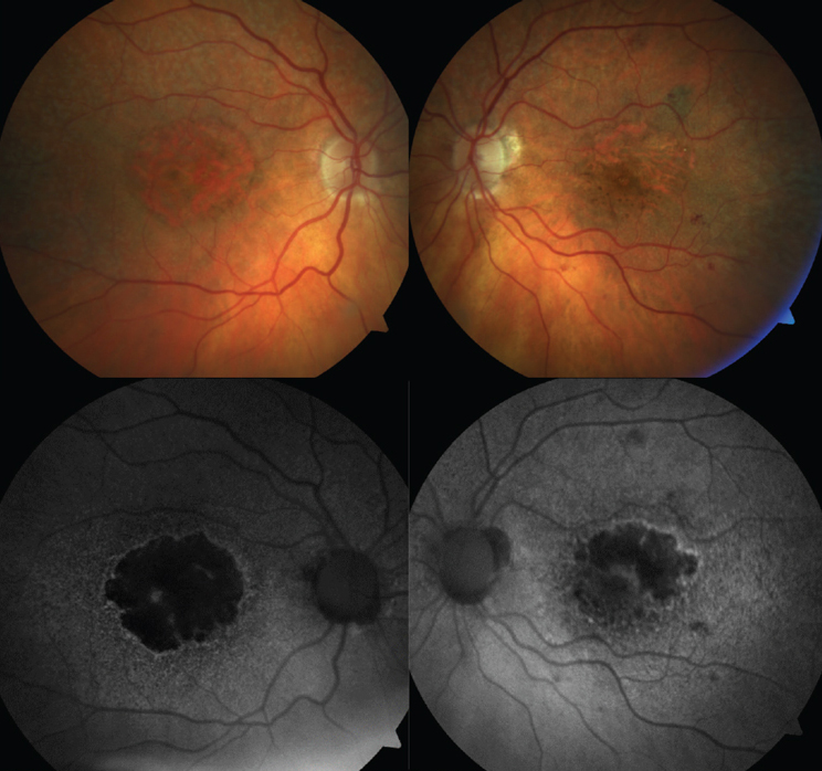
Geographic atrophy research has made several recent breakthroughs with the approval of a new complement-based therapy (pegcetacoplan) and another (avacincaptad pegol) expected to follow, but there’s still much that remains unclear about the mechanisms behind geographic atrophy and intermediate AMD progression. Identifying new, clinically meaningful early biomarkers of progression is a step forward, and researchers recently reported several that can be identified with good agreement by clinicians in a paper published in Ophthalmology Retina.
In the study (funded by Genentech), the researchers assessed the ability to evaluate progression of individual lesions of incomplete or complete retinal pigment epithelium (RPE) and outer retinal atrophy (iRORA/cRORA). Their analysis included second-eye OCT images from the multicenter, prospective Proxima B clinical trial (NCT02399072) cohort with geographic atrophy in one eye and iAMD in the other at baseline. OCT imaging was obtained according to a standardized protocol. Junior and senior readers analyzed the OCT images for nine qualitative features of early atrophy. Identified features were quantified. Inter-reader agreement for qualitative and quantitative assessments was recorded.
There was moderate agreement for presence of choroidal hypertransmission, followed by external limiting membrane disruption, outer plexiform layer/inner nuclear layer subsidence and a hyporeflective wedge-shaped band. The researchers wrote in their paper that these were “the most robust OCT features” for assessing progression of iAMD towards geographic atrophy by expert graders.
The highest agreement for quantitative measurements was for choroidal hypertransmission and the lowest was for RPE attenuation/disruption. Significant progression was seen with choroidal hypertransmission and external limiting membrane disruption, according to longitudinal adjudicated changes. Ellipsoid zone disruption and RPE attenuation/disruption did not show significant progression.
The researchers concluded in their paper that these findings indicate that many OCT features of iRORA and cRORA can be identified, annotated and quantified reliably. This “may be helpful in assessing new therapeutic interventions,” they wrote.
Schmitz-Valckenberg S, Saßmannshausen M, Braun M, et al. Inter-reader agreement and longitudinal progression of incomplete/complete retinal pigment epithelium and outer retinal atrophy in AMD. Ophthalmology Retina 2023. [Epub ahead of print]. |
