 |
Q: Given the recent outbreak of fusarium keratitis, how can I maximize the laboratory yield when I suspect this fungus in contact lens wearers?
A: There are five things you can do:
Correctly scrape the cornea. Fungus tends to hide under a bed of necrotic tissue. Therefore, the first thing you should do to maximize your yield is to scrape the leading edge of the ulcer and at the base where the greatest number of organisms should be present, says optometrist Arthur B. Epstein, chair of the AOAs Contact Lens & Cornea Section.
The appropriate scraping tool: a Kimura spatula or a Bard-Parker blade.
Culture the patients contact lens case, contact lenses and contact lens solution. Because fusarium can be elusive, you want to culture everything the patient uses that is related to his or her contact lens regimen, says ophthalmologist Dean Ouano, of New Bern, N.C.
Place the culture scraping in a media that does not have inhibitors. Many available media for culturing fungus contain medications to keep saprophytes, or useless junk, from cluttering the culture plate. This is done because fungus cultures are usually taken by pulmonary medicine professionals who are only interested in the severe kinds of fungus that can infect the lung, says ophthalmologist Herbert E. Kaufman, of Louisiana State University Medical Center School of Medicine in New Orleans.
Non-medicated media: Sabarouds agar plate, blood agar plate and thioglycolate broth. All can be obtained from your local hospital. Generally speaking, if you have enough of a yield, you can grow fungus species within 72 hours, Dr. Ouano says. You can store these culture media in your office refrigerator.
 |
 |
|
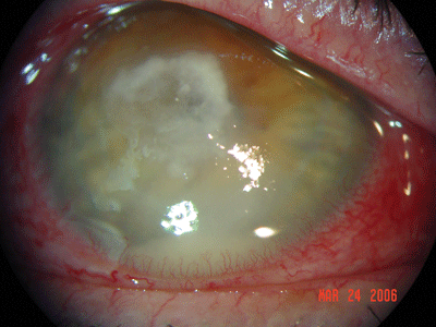
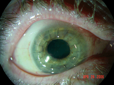
Top: Fusarium keratitis seen in a soft contact lens patient who used ReNu with MoistureLoc. This patient was misdiagnosed with contact-lens related cornea edema and iritis and was treated by her optometrist and referring ophthalmologist with Pred Forte every two hours. When the drops did not work, the patient was referred to a corneal specialist, who thought she had herpetic eye disease. Notice the white endothelial plaque, which is fairly inherent to fusarium. Bottom: The same patient six weeks after undergoing a corneal transplant. |
Use a potassium hydroxide (KOH) preparation stain. Place the ocular scrapings on a slide that contains KOH solution. Without heat, the solution will dissolve the skin cells but has no effect on the fungus cells. So, youll be able to see the fusarium, if it is present, Dr. Kaufman says.
Q: Has the standard of waiting for a positive culture result before treating suspected fusarium changed since this outbreak has been noted in contact lens wearers, especially in those who have used and are still using ReNu with MoistureLoc?
A: Practitioners may be tempted to begin treatment immediately, but the outbreak shouldnt change the standard of care, some experts say.
Indeed. Pharmacist Charles Leiter, of San Jose, Calif., says that requests for both natamycin and voriconozole have doubled since the reported fusarium outbreak. Weve increased the amount of natamycin we order, and instead of ordering five bottles of Natacyn from Alcon, weve been ordering 10 to keep up with the requests, he says.
Still, this rise in requests for and sales of anti-fungal medications may be due to the reported fusarium outbreak itself and not a change in the standard of care.
Regardless, do not treat suspected fusarium keratitis empirically. A patient who presents with an infiltrate and reports use of ReNu with MoistureLoc may not have fusarium keratitis. Rather, he or she may have a bacterial, viral or other infectious form of keratitis that is completely unrelated to this contact lens solution.
 |
 |
|
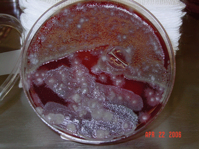
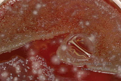
Blood agar culture plates teem with fusarium in this patient who used ReNu with MoistureLoc solution. Criteria that should lead you to culture the infiltrate include prior history of corticosteroid use; poor clinical response to anti-bacterial therapy; history of trauma with vegetative matter, such as a tree branch or soil; or prior use of ReNu with MoistureLoc. |
This is important because a misdiagnosis of fusarium keratitis leads to the wrong treatment. The wrong treatment can be detrimental to the patient in that antifungal medications are more toxic to the ocular surface and the cornea than standard antibacterial therapies.
Natamycin, for instance, can cause eye irritation, redness and swelling. So, you do not want to commit the patient to several weeks of treatment with this drug unless you are absolutely sure the patient has fusarium keratitis, Dr. Ouano says.
Also, natamycin is completely ineffective against viral, bacterial and protozoan infections. The result? A very unhappy patient who still has the infiltrate, plus the added complication of ocular surface problems stemming from the toxicity of the wrongly prescribed medication.
So, how should you manage a patient you suspect of having fusarium keratitis until the culture results return?
Prescribe one of the fourth-generation fluoroquinolones, namely Zymar (gatifloxacin, Allergan) or Vigamox (moxifloxacin, Alcon), before the culture results return. Why? Most corneal ulcers are bacterial in nature. Also, be sure to monitor the patient closely, Dr. Epstein says. If the culture results come back positive for fusarium, switch the patient to an anti-fungal medication immediately.
