A 91-year-old white female presented to the office last month as a new patient seeking a second opinion. She was concerned about her significantly decreased vision and inability to read. The patient was active and alert, noticing progressively decreasing vision O.U. over the past year. Her ocular history was remarkable for bilateral cataract surgery with posterior chamber IOL implantation in the late 1980s.

She was aware that she had bilateral macular degeneration and her last doctor told her that she had glaucoma, but didn’t need drops. This concerned her because she apparently has several friends with glaucoma—all of whom are currently using drops.
Her systemic medications included warfarin, lisinopril, isosorbide, atenolol and pravastatin. She reported no allergies to medications and denied a family history of glaucoma.
Diagnostic Data
Her entering visual acuities were finger counting at two feet O.D. and O.S. Pupils did not demonstrate an afferent papillary defect, although both were minimally reactive and irregular.
Extraocular muscles were full in all positions of gaze. Confrontation fields were severely constricted O.U.
Her visual acuity did not improve through myopic or astigmatic correction. While reading the chart, she typically viewed eccentrically.
A slit lamp examination of her anterior segments was remarkable for bilateral, limbal-based cataract incisions with surgical peripheral iridotomy bilaterally. Both irides were characterized by sectoral atrophy and pupillary ruff damage, probably occurring during cataract surgery. Her corneas were clear O.U., and anterior chambers were deep and quiet O.U.
Applanation tensions were 14mm Hg O.D. and 15mm Hg O.S. Central corneal thickness (CCT) readings measured 558μm O.D. and 570μm O.S.
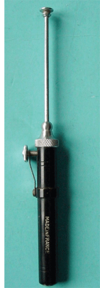
When using this instrument, you are decreasing perfusion through the central retinal artery, with the intent of measuring blood pressure in these arteries.
Through dilated pupils, her implants were well centered in the capsular bags, and both posterior capsules were opened. Both IOLs were pitted on the posterior surface, consistent with YAG nicks.
Close examination of her optic nerves demonstrated moderate glaucomatous cupping measured at 0.75 x 0.90 O.D. and 0.80 x 0.90 O.S. What was left of her neuroretinal rims appeared pink and moderately perfused. The cups were vertically elongated and the neuroretinal rims were notched inferotemporally O.D. and O.S. The superior temporal neuroretinal rims were also markedly thinned. There was a moderate amount of peripapillary atrophy (PPA) O.U. The patient’s vascular examination was characterized by moderate arteriolar attenuation, hypertensive retinopathy grade 1 O.U. and arteriolarsclerotic retinopathy grade 2 O.U. with crossing changes. Both maculae were characterized by geographic retinal pigment epithelium (RPE) atrophy; there was no evidence of subretinal fluid or leak in either eye. There were bilateral posterior vitreous detachments (PVD), and her peripheral retinal examinations were unremarkable.
While the patient was dilated, digital retinal and stereo optic nerve images were obtained.
Currently, the patient is scheduled for a full glaucoma work-up, including threshold visual fields, repeat IOP measurement, gonioiscopy and HRT-3 imaging of the optic nerves, at which time she will ultimately be medicated for glaucoma.
I explained that, due to my findings, we would not be able to improve her vision with traditional means. We also discussed the need for a low vision evaluation and anti-glaucoma medication therapy.
Discussion
This very active and alert 91-year-old patient suffers from visual impairment from both non-angiogenic AMD as well as advanced glaucoma. We are waiting for the results of visual field studies, so I cannot comment precisely on her field status. However, after looking at her posterior pole photos, we can clearly see the effects to her central vision from AMD.
Nonetheless, considering her significant neuroretinal rim loss and constricted confrontation fields, I would postulate that she also in fact has moderate field loss attributable to the untreated glaucoma.
What is disconcerting about this case is that she apparently was told that she had glaucoma, but was not medicated. Perhaps her pervious eye doctor felt as though the costs of initiating treatment outweighed any possible benefit, given her age. However, she had been seeing the same providers for 20+ years, and during that time, there should have been some documented changes over time. Be that as it may, she is not medicated, and needs to be medicated.
But the question lingers: Why no treatment previously? She clearly has “normal” IOP, average CCT measurements and compromised neuroretinal rims.
I speculate that the PPA causing a “blanching” of the optic nerve and perioptic appearance may have played a role. In some cases—especially with high myopes with PPA—it can be difficult to actually assess the neuroretinal rim characteristics carefully, especially in the context of glaucoma. But when you look closely at her neuroretinal rims, you can easily discern the optic canal, a thinned neuroretinal rim and the PPA. I think this patient had progressive normal tension glaucoma that was simply missed. Consider this when you do a cursory look at the optic nerve and see normal pressures and CCT measurements.
It is well documented that elevated IOP is a significant risk factor for the development and progression of glaucoma. For non-pressure dependent glaucoma, our attention turns to thoughts of decreased perfusion to the optic nerve. Especially in an elderly patient with a history of cardiovascular disease, it is not unusual to expect decreased perfusion. Given this patient takes blood pressure medications and shows vascular retinal attenuation, she may have compromised blood flow—especially at night—when blood pressure tends to drop in both healthy and hypertensive patients. Recent studies confirm that lowering blood pressure, especially at night, can compromise the optic nerve.1-3
Ocular perfusion pressure (OPP) refers to the blood pressure supplying the optic nerve, retina and perhaps the choroid. OPP is affected by both blood pressure and IOP as well as the regulation of ocular blood flow via a process called autoregulation. It is important to note that retinal blood flow and pressure is more subject to autoregulation than is choroidal circulation. It is also important to remember that perfusion to the optic nerve head is via the posterior ciliary arteries, while perfusion to the retinal ganglion cells is via the retinal arteries.
Current research is looking to elucidate the precise relationships between OPP, blood pressure, IOP and maintaining nerve integrity. Ocular blood flow analyzers are now available clinically, and there are a few formulae currently being discussed for estimating retinal blood pressure by measuring systemic blood pressure (usually measured at the arm) and IOP.
But, is there an easy and timely way to more directly measure retinal arterial systolic and diastolic blood pressure without expensive instrumentation? It is time to look to an instrument that was used 20 to 30 years ago—the ophthalmodynomometer. A Balliart ophthalmometer is an instrument that is used in conjunction with ophthalmoscopy to measure retinal arterial systolic and diastolic blood pressure. When done properly, these measurements can be obtained in a matter of seconds. It works on the principle of evaluating the pressure at which retinal arterial pulsation and collapse occurs. When IOP is elevated to the level just above retinal arterial diastolic pressure, retinal arteries begin to pulsate. But when IOP is raised further to the level of retinal arterial systolic pressure, the retinal arteries collapse.Though it may not be prudent to temporarily compromise retinal artery flow in a case of advanced normal tension glaucoma, this measurement can be obtained very quickly.
Perhaps we might want to get an estimate of retinal blood pressure in our younger normal tension glaucoma suspects before they become elderly with a significant amount of neuroretinal rim compromise and vision loss. Just a thought.
1. Venkataraman ST, Flanagan JG, Hudson C. Vascular reactivity of optic nerve head and retinal blood vessels in glaucoma--a review. Microcirculation. 2010 Oct;17(7):568-81
2. Krasinska B, Karolczak-Kulesza M, Krasinski Z, et al. A marked fall in nocturnal blood pressure is associated with the stage of primary open-angle glaucoma in patients with arterial hypertension. Blood Press. 2010 Dec 7. [Epub ahead of print]
3. He Z, Vingrys AJ, Armitage JA, Bui BV. The role of blood pressure in glaucoma. Clin Exp Optom. 2011 Mar;94(2):133-49.
