 |
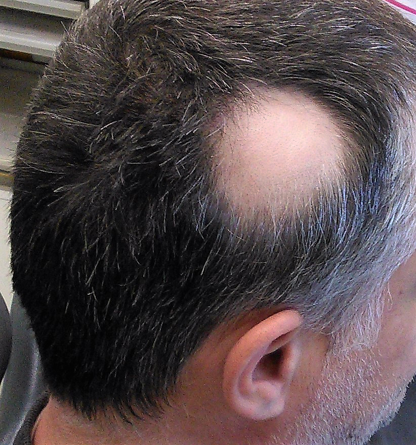
| Melanocytes in the choroid may be the link to the observed inflammation, as alopecia is mediated by T lymphocyte activity against pigmented cells. Photo: Thirunavukkarasye-Raveendran/Wikimedia Commons. Click image to enlarge. |
The pathogenesis of alopecia areata (AA) is unclear, but research suggests its development may involve an autoimmune mechanism mediated by T-cells. In some AA cases, ocular abnormalities such as cataract, tear film disruption and chorioretinal changes have been reported, leading researchers to recently investigate how retinal layers and choroidal structures may be affected in these individuals. Their study found that the following may be observed in AA patients: T lymphocyte-mediated hair follicle damage, choroidal melanocyte damage and inflammation. They also concluded that choroidal thickness may increase secondary to melanocyte inflammation in these patients.
The study examined the right eyes of 42 AA patients and 42 controls through an ophthalmic examination and SD-OCT. The researchers observed that in alopecia patients, choroidal thickness was significantly greater in the subfoveal, temporal and nasal regions compared with the control group.
The average thickness of the following layers was not different between alopecia patients and controls: ganglion cell layer, inner plexiform layer, inner nuclear layer, outer plexiform layer, outer nuclear layer, retinal pigment epithelium (RPE), inner retinal layers and photoreceptor layers.
“Considering that the main pathophysiology in AA is T lymphoid-mediated inflammation against melanocytes, the fact that we were unable to identify any significant difference in thickness between the macula and retinal nerve fiber layer (RNFL) can be attributed to the absence of melanocytes in the macula and RNFL,” the researchers explained in their paper on the study findings. While the RPE does contain melanocytes, the authors hypothesized that the lack of a difference between RPE thickness in patients and controls may be attributed to the amount of melanin pigment and its position and distribution in RPE, as prior research has shown that RPE-related melanosomes may become displaced under various light frequencies (i.e., on OCT images).
The researchers conclude that due to their thicker choroids, patients with alopecia should be closely monitored for possible posterior ocular segment disorders.
Oren B, Aksoy Aydemir G, Duzayak S, Kiziltoprak H. Evaluation of retinal layers and choroidal structures using optical coherence tomography in alopecia areata. Medeni Med J. 2023;38:140-7. |
