 Februarys column covered the role of nutritional supplements in preventing age-related macular degeneration (AMD). In it, we talked about the growing body of evidence, which suggests that various nutritional supplements and antioxidants, such as carotenoids, have a protective effect against AMD.1-5
More specifically, other studies have demonstrated an inverse correlation between high intake levels of certain carotenoids, such as lutein and zeaxanthin, and the risk of developing AMD.6,7
Lutein and zeaxanthin are distinguished from other caretonoids because they are found in high concentrations within the macula. Research indicates that lutein and zeaxanthin minimize oxidative stress on the macula by absorbing blue light and neutralizing free radicals.8
The protective macular pigment (MP) is comprised primarily of lutein and zeaxanthin.9 So, low levels of MP may be associated with an increased risk for AMD. Here, well describe how you can protect your patients against AMD by measuring and improving their macular pigment optical density (MPOD) scores.
Measuring MPOD
Detection of low MPOD levels is now possible. MPOD is evaluated through the use of non-invasive psychophysical measurements.10-12 The most widely used technique is based on the principles of heterochromatic flicker photometry (HFP), the comparison of luminosity between two alternating wavelengths. Previous HFP devices required the patient to adjust the brightness of one of the lights until the flicker was reduced to a minimum.13
Current commercially available instruments used to assess MPOD include QuantifEYE (ZeaVision, LLC) and MacuScope (MacuChek).14,15 The QuantifEYE and MacuScope are scientifically validated devices that measure MPOD by simply requiring the patient to press a button in response to the appearance of flicker.14 The test is quick, does not require dilation and is typically unaffected by ocular media.
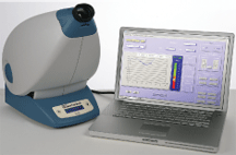
QuantifEYE (with laptop display) measures a patient"s MPOD and can help identify individuals at risk for AMD.
Image courtesy: ZeaVision, LLC
The instrument presents the patient with two alternating lights of different wavelengths: blue light, which is absorbed by the MP, and green light, which is outside the absorption of MP. A series of alternating lights flash at varying frequencies and brightness intensities, resulting in the patients perception of flicker.
The test is typically performed centrally in the fovea, where the highest concentration of MP exists, and parafoveally (eccentrically located 6 to 8 from the central fovea).14,15 This eccentric location correlates to an area where there is absence of MP. For some devices, a sensitivity test precedes both the central and peripheral tests.14 The sensitivity test is determined by the initial five responses to flicker. Depending on the sensitivity to flicker, white light is added to either the green or blue light.
Since age-related ocular media can contribute to increased yellow pigment in the periphery, the QuantifEYE instrument determines an estimated peripheral MPOD value based on the patients age.14 So, performing the actual peripheral MPOD test is only recommended for patients with a history of diabetes, cataract, macular degeneration or cataract surgery.14
The MPOD test is reportedly accurate and replicable, but because it is a psychophysical test, it is prone to human error. It is a subjective test that requires the patients full cooperation. In order to add precision and reliability, the QuantifEYE displays data as spectral absorption curves for both the central and peripheral test.14 Since we expect to see a specific graph pattern, we can easily determine if the test was conducted accurately. Thorough evaluation of these characteristic graph patterns helps verify the accuracy of the MPOD score.
The patients MPOD score is the ratio of the central blue light absorption rate to the peripheral absorption rate.14-16 This score is compared to a normative database, and the patient is placed into either the low-risk or high-risk category. The more blue light is absorbed, the higher the MPOD is.13 Patients with lower levels of MPOD are exposed to more harmful blue light, increasing their risk of developing AMD. These patients are at greatest need of nutritional supplementation.
Who Should Be Screened?
These instruments are not designed for patients who already have advanced AMD. Rather, adults with known risk factors typically benefit the most from MPOD measurement. However, since high MPOD is correlated with decreased progression of AMD, patients with early signs of the condition would also benefit from this test.10,17
If you can identify a patient who is at risk for AMD associated with lower MPOD levels at an early stage, you will be able to intervene to slow disease progression. In such cases, you should recommend nutritional supplementation to increase the individuals MPOD.
Benjamin Franklin once said, An ounce of prevention is worth a pound of cure. This statement takes on new meaning in the era of technological advancement in AMD management. Instruments that measure MPOD may help identify your patients who are at risk for developing AMD. Early detection and treatment are critical in mitigating irreversible vision loss.
1. Chong EW, Wong TY, Kreis AJ, et al. Dietary antioxidants and primary prevention of age related macular degeneration: systematic review and meta-analysis. BMJ 2007 Oct 13;335(7623):755.
2. Christen WG,
3. Seddon JM, Ajani UA, Sperduto RD, et al. Dietary carotenoids, vitamins A, C, and E, and advanced AMD: Eye Disease Case-Control Study Group. JAMA 1994 Nov 9;272(18):1413-20.
4. Age-Related Eye Disease Study Group. A randomized, placebo-controlled, clinical trial of high-dose supplementation with vitamins C and E, beta carotene, and zinc for age-related macular degeneration and vision loss: AREDS report no. 8. Arch Ophthalmol 2001 Oct;119(10):1417-36.
5. Cangemi FE. TOZAL Study: an open case control study of an oral antioxidant and omega-3 supplement for dry AMD. BMC Ophthalmol 2007 Feb 26;7:3.
6. Mares-Perlman JA, Fisher AI, Klein R, et al. Lutein and zeaxanthin in the diet and serum and their relation to age-related maculopathy in the third national health and nutrition examination survey. Am J Epidemiol 2001 Mar 1;153(5):424-32.
7. Goldberg J, Flowerdew G, Smith E, et al. Factors associated with age-related macular degeneration. An analysis of data from the first National Health and Nutrition Examination Survey. Am J Epidemiol 1988 Oct;128(4):700-10.
8. Stahl W, Sies H. Physical quenching of singlet oxygen and cis-trans isomerization of carotenoids. Ann N Y Acad Sci 1993 Dec 31;691:10-9.
9. Snodderly DM. Evidence for protection against age-related macular degeneration by carotenoids and antioxidant vitamins. Am J Clin Nutr 1995 Dec;62(6 Suppl):1448S-1461S.
10. Richer S, Stiles W, Statkute L, et al. Double-masked, placebo-controlled, randomized trial of lutein and antioxidant supplementation in the intervention of atrophic age-related macular degeneration: the Veterans LAST study (Lutein Antioxidant Supplementation Trial). Optometry 2004 Apr;75(4):216-30.
11. Mellerio J, Ahmadi-Lari S, van Kuijk F, et al. A portable instrument for measuring macular pigment with central fixation. Curr Eye Res 2002 Jul;25(1):37-47.
12. Loane E, Stack J, Beatty S, Nolan JM. Measurement of macular pigment optical density using two different heterochromatic flicker photometers. Curr Eye Res 2007 Jun;32(6):555-64.
13. Bone RA, Landrum JT. Heterochromatic flicker photometry. Arch Biochem Biophys 2004 Oct 15;430(2):137-42.
14. ZeaVision, LLC. ZeaVisions QuantifEYE Program: First of its kind for assessing risk of developing age-related macular degeneration (AMD). Available at: www.zeavision.com (Accessed February 2008).
15. MacuChek. MacuScope. Available at: www.macuscope.com (Accessed February 2008).
16. Sharpe LT, Stockman A, Knau H, Jgle H. Macular pigment densities derived from central and peripheral spectral sensitivity differences. Vision Res 1998 Nov;38(21):3233-9.
17. Bone RA, Landrum JT, Cao Y, et al. Macular pigment response to a supplement containing meso-zeaxanthin, lutein and zeaxanthin. Nutr Metab (Lond) 2007 May 11;4:12.
