 |
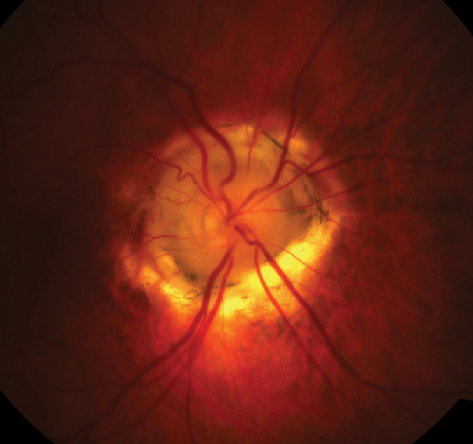
| MGDA may cause an irregular optic nerve pattern in patients. Photo: Nevi Hehar, OD. Click image to enlarge. |
The morning glory disc anomaly (MGDA) is a rare congenital malformation of the optic disc. Usually unilateral and sporadic, MGDA is diagnosed clinically through fundoscopy. It is characterized by a funnel-shaped posterior pole, including an enlarged and malformed optic disc, circumscribed by a peripapillary pigmented ring, and sometimes contractile smooth muscle fibers, and covered by the central glial tissue, whereas retinal vessels emerge from the periphery of the disk in a radial pattern. Its association with a significant enlargement of the optic nerve has been recently reported in a few cases raises the question of potentially associated optic nerve gliomas. A retrospective study of all cases of MGDA in a hospital in Paris between 2008 and 2018 revealed that, while some characteristics were compatible with glioma, there were no changes in the optic nerve thickness during follow-up.
The study included nine individuals (six females and three males) who all presented with unilateral MGDA without dysmorphic systemic features. The median age at MGDA diagnosis was 14 months. The main sign having led to MGDA diagnosis was convergent strabismus. The median age at first MRI imaging was 3.8 years.
MRI showed posterior protrusion of the globe (staphyloma) centered by the optic disc in all cases, and ipsilateral optic nerve abnormalities were also found in all cases. The optic nerve was thinner than the contralateral one in its intraorbital, intracanalar and intracranial portions in one case (11%). In eight cases (89%), the thickness of the optic nerve was irregular and varied along its pathway: thick, normal and/or thin. The contralateral optic nerve and hemichiasm in these cases were always normal.
On MRI, some characteristics were compatible with optic nerve glioma features, notably irregular, tortuous optic nerves. When gadolinium injection had been performed (three cases), the researchers found that none exhibited gadolinium enhancement. When serial MRI scanning was available (four cases), there was no evolution of the abnormalities with a follow-up of 3.5 years.
“Such a pattern of irregular, moniliform optic nerves is unique and could represent an MRI criterion of morning glory disc syndrome, useful in case of a doubtful optic disc appearance on fundus examination,” the researchers noted.
“In patients with MGDA, optic nerve and chiasm abnormalities are the rule, with most often a unique pattern of irregular optic nerve thickness—hypertrophy and hypoplasia—from the orbit to the chiasm, they concluded. “Such pattern should be recognized and points to a developmental abnormality, rather than an optic nerve glioma.”
Nguyen DT, Boddaert N, Bremond-Gignac D, Robert MP. Optic nerve abnormalities in morning glory disc anomaly: an MRI study. J Neuroophthalmol. November 18, 2021. [Epub ahead of print]. |
