 A 55-year-old white female presented with no ocular or visual complaints. During the dilated fundus examination, we observed a choroidal lesion. The lesion measured 4.0 disc diameters (DD) in size, appeared flat, and was associated with numerous overlying drusen deposits (figure 1). We noted no other pertinent findings.
A 42-year-old white female presented for an annual comprehensive eye exam. Dilated fundus examination revealed a 2.5DD choroidal lesion. The lesion appeared elevated and had an overlying orange pigmentation (figure 2). Optical coherence tomography revealed the presence of subretinal fluid. We detected no associated drusen or halo nevus.
Although both patients exhibit somewhat similar choroidal lesions, one presentation has the potential to be cancerous. But how can you effectively determine which lesion that is?
Nevi vs. Melanomas
Choroidal nevi and choroidal melanomas have many overlapping features, which poses a diagnostic dilemma. Yet, several distinguishable characteristics may be noted in both lesions.
• Choroidal nevi are relatively common, and are documented in 5% to 10% of the American population.1 They are flat, green or slate gray choroidal lesions that typically measure less than 3.0DD, yet the size and thickness of choroidal nevi vary widely. Associated findings include serous retinal pigment detachment, choroidal neovascularization pigmentary changes and drusen formation.2
• Choroidal melanomas are the most common ocular malignancy. They appear as darkly pigmented lesions and typically present with some degree of elevated thickness. Choroidal melanomas are classified as small (<10mm in diameter and <3mm thickness), medium (10mm to 15mm in diameter and 3mm to 5mm in thickness) or large (>15mm in diameter and >5mm in thickness).3 Features may include lipofuscin and associated serous fluid.4 Documented evidence of short-term growth seems to be a pathognomonic sign of a choroidal melanoma.5 And although some studies have shown progressive enlargement of choroidal nevi, malignant features are absent.6 Whether melanomas arise from pre-existing nevi or are completely separate entities is somewhat debatable.
Small Melanoma Characteristics
Typically, patients with larger, thicker melanomas have worse long-term prognoses than individuals with smaller lesions. Several meta-analyses indicate that patients with choroidal tumors exhibit a five-year mortality rate that ranges from 15% to 50%, depending upon lesion size and thickness.4,7
Therefore, early detection of choroidal melamomas is crucial to minimize the risk of metastatic disease. A prompt diagnosis and timely referral for proper management will increase the patient’s overall chances for survival dramatically.7
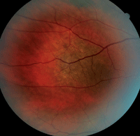
1. Fundus photograph of our first patient. What do you notice?
Because histological confirmation of a choroidal melanoma is not feasible, we chiefly rely on clinical characteristics of growth to determine the risk of metastasis. Within the last 15 years, Carol L. Shields, M.D., and associates conducted two retrospective medical reviews of patients with presumed small choroidal melanomas.4,8 In both studies, the authors’ primary goal was to identify specific features associated with small choroidal melanoma that could predict lesion growth and potential metastasis.
In the first study, the researchers evaluated the records of approximately 1,300 patients who were followed regularly from 1970 to 1990.4 They determined that the average diameter of the subjects’ choroidal lesions was 5mm, with thicknesses ranging from flat to 3mm. All patients underwent standardized testing, including dilated fundus examination and ultrasonography.
At the study’s conclusion, the authors identified five common traits associated with small choroidal melanomas:4
- Thickness (>2mm)
- Fluid (retinal)
- Symptomatic (photopsia, decreased visual acuity, metamorphopsia, etc.)
- Orange pigment (lipofuscin)
- Margin near the nerve (within 3mm)
These features may be remembered more easily using the mnemonic “To Find a Small Ocular Melanoma.”
In the second study, Dr. Shields and associates sought to determine whether additional clinical characteristics might be indicative of a risk of malignancy.8 This retrospective medical review included nearly 2,500 patients followed from 1974 to 2006. At the study’s completion, the researchers documented the presence of two additional features:8
- Ultrasonography hollowness
- Halo nevus
This discovery adds to the mnemonic: “To Find a Small Ocular Melanoma, Use Helpful Hints.”
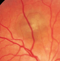
2. Fundus image of our second patient. How does this presentation differ from the findings observed in our first patient?
The researchers noted that patients with three or more of the traits had a 50% higher risk of melanoma growth.8 Further, they determined that patients with three to four traits were 15% to 20% more likely to experience metastasis; those with one to two factors were at a 5% higher risk of metastasis; and individuals with zero risk factors had less than a 1% chance of metastasis.8
Accordingly, a management protocol was developed for these small choroidal melanomas, based on the number of documented traits:
- 0: Patients should be followed on an annual basis.
- 1-2: Patients should be followed every four to six months to evaluate both short- and long-term lesion growth.
- 3 or more: Patients should be referred to a specialist for further management and treatment.
Back to Our Patients
Given the overall clinical features of our patients introduced above, we diagnosed the first individual with a large choroidal nevus and the second individual with a small choroidal melanoma. Malignant features in the second case included lesion thickness as well as the presence of orange lipofuscin and associated serous fluid.
Our diagnostic accuracy when encountering small choroidal melanomas continues to improve. Early recognition of high-risk characteristics helps the clinician make a prompt referral, which ultimately translates into a better prognosis for the patient.
1. Sumich P, Mitchell P, Wang JJ. Choroidal nevi in a white population: the Blue Mountains Eye Study. Arch Ophthalmol. 1998 May;116(5):645-50.
2. Gonder JR, Augsburger JJ, McCarthy EF, Shields JA. Visual loss associated with choroidal nevi. Ophthalmology. 1982 Aug;89(8):961-5.
3. The Collaborative Ocular Melanoma Study (COMS) randomizedtrial of pre-enucleation radiation of large choroidal melanoma II: initial mortality findings. COMS report no. 10. Am J Ophthalmol. 1998 Jun;125(6):779-96.
4. Shields CL, Shields JA, Kiratli H, et al. Risk factors for growth and metastasis of small choroidal melanocytic lesions. Ophthalmology. 1995 Sep;102(9):1351-61.
5. Thiagalingam S, Wang JJ, Mitchell P. Absence of change in choroidal nevi across 5 years in an older population. Arch Ophthalmol. 2004 Jan;122(1):89-93.
6. Elner VM, Flint A, Vine AK. Histopathology of documented growth in small melanocytic choroidal tumors. Arch Ophthalmol. 2004 Dec;122(12):1876-8.
7. Markowitz JA, Hawkins BS, Diener-West M, Schachat AP. A review of mortality from choroidal melanoma. I. Quality of published reports, 1966 through 1988. Arch Ophthalmol. 1992 Feb;110(2):239-44.
8. Shields CL, Furuta M, Berman EL, et al. Choroidal nevus transformation into melanoma: analysis of 2514 consecutive cases. Arch Ophthalmol. 2009 Aug;127(8):981-7.
